Case Study of Amniotic Fluid Embolism in Obstetrics: Diagnosis and Management
380 likes | 553 Vues
Learn about a complex obstetric disorder, Amniotic Fluid Embolism (AFE), its onset, predisposing factors, and clinical manifestations in this detailed case study from Xiong Yu Obstetric & Gynecology Hospital at Fudan University.

Case Study of Amniotic Fluid Embolism in Obstetrics: Diagnosis and Management
E N D
Presentation Transcript
Amniotic Fluid Embolism Xiong yu Obstetric & Gynecology Hospital, Fudan University
Case 1 • Gravida 3 and Parturition 1,41w, labor induced by propess, membrane ruptured naturally, at the same time, dyspnea and cyanosis occurred. • What happened? If you are the doctor,what should you do? • Trachea cannula was done immediately, and CS was prepared. • The baby was survival with 9 apgar score. • In the operation, sudden cardiac arrest occurred and autonomously heartbeat recovered after 50 minutes external chest compression. • But DIC emerged, hysterectomy was done. Finally, the patient’s life was saved,but she became a vegetable. • One year later, she died.
Amniotic Fluid Embolism(AFE) • Serious intrapartum complication • A complex disorder caused by amniotic fluid entering maternal circuration and classically characterized by the abrupt onset of hypotension, hypoxia, and consumptive coagulopathy • Incidence:1:20000 • Mortality: 80%, in term pregnancy
Condition of AFE Onset • Rupture of membrane • Hypertonia of amnion cavity • Open blood sinus • Injury of cervical canal or uterine wall • Placenta previa, placenta abruption, placenta marginal sinus rupture • Adherence site of placenta
Predisposing factors • Premature rupture of membrane, artificial rupture or stripping of membrane, artificial expansion of cervix • Too strong uterine contraction • Rigidity contraction and precipitate labour caused by inappropriate using of oxytocin and operation in cavity • Injury • Cervical laceration, rupture of uterus, uterine incision in caesarean section,forcep curettage
Some pathological pregnancy • Twin, multiplets, macrosomia, polyhydramnions, prolonged labour, dystocia, placenta abruption, placenta previa, retention of dead fetus, infection of amnion cavity, fetal distress • Press abdoman and uterus by brute force
Pathway of amnion fluid entering maternal blood circulation • Internal cervical vein Amniotic fluid volume entering maternal circulation related to : strength of contraction degree of injury • Uterine placenta bed : • Broken venule in adherence site of placenta • Fissure in adherence site of placenta • Open decidua blood sinus • placenta marginal vessel • Amniotic permeationpressure of amniotic cavity↑intensity of amniotic membrane ↓
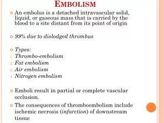
Pathyphysiology Pulmonary hypertension Allergic shork Disseminated intravascular coagulation(DIC) Acute renal failure
Amniotic fluid (Epithelial cell, mucus,meconium, vernix caseosa, lanugo) Ⅰallergic reaction Maternal circulation reflectively pulmonary circulation Vessel embolism Vessel spasm bronchospasm bronchi secretion increased Bleeding without coagulation chill bronchiostenosis Vessel block ,stenosis ventilation obstruct Pulmonary hypertention toxicosis and anoxia of the whole body returned blood volume to LA output Acute respiratory failure Acute pulmonary heart disease Histanoxia (cyanosis) renal anoxia cerebral anoxia, Anxious, seizure, coma Respiratory acidosis Right heart failure Acute renal failure Metabolic acidosis Peripheral circulatory failure, Bpdrop shork
Clinical manifestation • Abrupt onset , critical oncoming force • Three stages appear in sequence in typical cases • Only mass vaginal bleeding and shork in atypical cases ——Degree of syptoms related to amount of particle matter in amniotic fluid, amount and velocity of amniotic fluid entering maternal circulatin
Premonitory symptom: Short-period dysphoria, chill and shiver, cough and dyspnea, cyanosis, vomit at the time of rupture of membrane • Symptoms disappear after treatment in mild AFE, severe AFE arise three stages : • Respiratory and circulatory failure, shork • Bleeding caused by DIC • Acute renal failure
Clinical manifestation ——the first stage • Acute respiratory and circulatory failure • Obvious cyanosis • Dyspnea • Coughing frothy sputum, raised heart rate, moist rales in lung • Fall of blood pressure • Coma, seizure • Severe cases: scream, respiratory arrest, cardiopulmonary arrest ,die
Clinical manifestation ——the second stage • Coagulation disorders hypercoagulable state→hypocoagulable stage • Bleeding of skin, mucosa, needle eye, incision • Hematuria, hematemesis • Mass vaginal bleeding • Typical symptom of DIC
Clinical manifestation ——the third stage • Multiple organ failure (MOF) • Acute renal failure • Oliguriaurine volume < 400ml/24h or 17ml/h • Anuria<100ml/24h ,metabolic disturbance • Azotemia • Obvious jaundice, ascite
Dignosis After rupture of membrane , after birth, or in operation Break out shiver, bucking, dyspnea, dysphoria, scream, cyanosis, seizure, bleeding, shork unkown reason, AFE should be considered Rescue immediately ——The key to improve rescue livability is correct and prompt dignosis, effective therapeutic measures
Assistant examination • Blood smear to find amniotic visible particle • Bedside chest X-lay • Bedside ECG or CDFI • Right atrium enlargement, cardiac damage • Laboratory examination related to DIC
Blood smear to find amniotic visible matter • Left A (case1)shows pink and wedge-shaped fetal squamous epithelium , left B(case2) shows pink fetal squamous epithelium surrounded by platelet • Right A(case1),right B(case2) showed fetal squamous epithelium from bronchic asearse fluid (red arrow)
Squamous epithelium in a peripheral pulmonary artery Fetal keratin in a peripheral pulmonary artery
Bedside chest X-lay • 70% of patients may have mild symptoms of lung edema • Disseminated effusion in pulmonary alveolus • Increased heart shadow
Coagulation examination • Dissipative hypocoagulability • Progressive drop of platelet count (<100×109/L) • Prothrombin time (PT >3”) • Kaolin active partial thromboplastin time( KPTT > 10”) • Fibrinogen < 1.5/L
Secondary hyperfibrinolysis • Plasma Protamine para-coagulation test (3Ptest) • Others • D-dimer • Antithrombase Ⅲ(AT Ⅲ) • Fibrinopeptide A(FDA) • Fibrin degradation production (FDP) • Capillary hemolysis • Broken RBC more than 10% in 20~30% blood smears of late stage DIC
Dignosis after death • To draw right ventricle blood for precipitation test to find visible particle of amniotic fluid • Autopsy • Notable right ventricle expansion • Pulmonary edema, alveolar hemorrhage , embolus containing amniotic particle in kidney, heart, brain, uterine, or broad ligament , embolus containing • Deciduous squamous epithelial cell from fetal skin • Lanugo • Fragment of fetal skin and amnion • Mucin from fetal intestinal tract • Bile from meconium
Differential diagnosis Air embolism:severe chest and back pain, sence of precordia pressure, occurred in rupture of uterus, placenta previa, operation in cavity Pulmonary embolism by thrombus:varicose vein and thrombophlebitis of lower limb, occurred at 9-14 days after birth, acute chest pain, bloody sputum, chest fricative, pulmonary embolism in X-ray Eclampsia:hypertention, proteinuria, shork appeared later Rupture of uterus:cephalopelvic disproportion, signs of impending of rupture of uterus ( abdominal pain, hematuria)
Prevention Reasonable using of oxytocin, to master indication and controll dose, watched by special person Avoid inappropriate operation in uterine cavity and birth canal injury Notice in artificial rupture of membrane Avoid pressing abdomen and uterus strongly at the time of delivery of baby Master indication of CS strictly Using sedative to suppress excessive contraction
Principle of management • To rescue quickly and decisively • To treat respiratory and circulatory failure firstly • Appropriate obstetric management
Step 1 Improve hypoxemia • Semireclining position • Oxygen uptake • High concentration oxygen(>50%) by mark,flow velocity 5-10L/min • Continious positive airway pressure by trachea cannula • Antiallergic • Dexamethasone 20mg iv, 20mg ivgtt p.r.n • Hydrocortisone 1000- 2000mg/d ivgtt
Relieve pulmonary hypertension • Paraverine : • Relax vascular smooth muscle • 30-90mg+5%GS20mlivgtt • Amniophylline : • Dilate coronary artery and bronchi smooth muscle • 250mg+5%GS 20ml ivgtt • Atropine : • Relieve pulmonary vasospasm, bronchospasm, cardiac depression • 1-2mg im or iv • Phentolamine : • Relieve pulmonary vasospasm • 5-10mg+5%GS100mlivgtt,adjust infusion rate according blood pressure
Step 2 Correct shork • Circulatory support with blood and component replacement : central venous pressure(CVP): 8-10cmH2O • Adjust vessel tensity: • dopamine 10-20mg+5%GS 250ml ivgtt • Treat acidosis: • 5%NaHCO3 100-200mlivgtt
Step 3 Treat DIC • Hypercoagulability in early stage——heparin 0.5mg-1mg/kg(heparin 1mg to be equivalent to 125IU) • First 25mg+NS100ml ivgtt in 1h • Then 25mg+5%GS500ml ivgtt • Clotting time maintain at 15min • Excessive heparin detoxified using 1% equivalent protamine solution • To plan CS is a contraindication of using heparin
Hyperfibrinolysis • Antifibrinolysis • 6-aminoacetic acid (EACA)ivgtt • P-aminiomethyl beozonic acid(Pamba)200-300mg/d ivgtt • Blood coagulation factors supply • Fresh blood • Fresh frozen blood plasma, condensation sediment • Platelet suspension , fibrinogen • VitK 20-40mg to promote liver to synthesis coagulation factors
Step 4 • Prevent heart failure: • Lanatoside 0.4mg+5%GS 20ml ivgtt slowly • Energy mixture • Prevent renal failure: • Furosemide 40mgiv,repeated p.r.n • Prevent infection: • To select broad-spectrum antibiotic with less renal toxicity
Obstetric management • Onset in first stage of labor ——termination of pregnancy by CS • Onset in second stage of labor ——termination of labor by vaginal midwifery • PPH occurred and not stopping bleeding ——hysterectomy