Download
1 / 40
410 likes | 497 Vues
Learn about signs, symptoms, and management of fetal distress during pregnancy and labor. Understand the importance of detecting abnormal fetal heart rate and meconium-stained amniotic fluid. Get insights into different types of decelerations and factors leading to fetal distress.

E N D
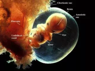
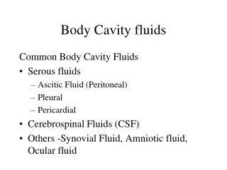
Fetal Distress • The term fetal distress is commonly used to describe fetal hypoxia during ante partum or intrapartum period. Which can result in fetal damage or death if it is not reversed or the fetus is not promptly delivered.
Signs Fetal distress can be detected via :- • Abnormal fetal heart ( fetal heart <100/min or >180 beat/ min ) • Thick meconium stained amniotic fluid • Abnormal cardiotocography ( Non reassuring fetal status) -Fetal tachycardia or bradycardia especially during & after contraction -Decreased beat-beat variability in base line fetal heart - Late deceleration
Biochemical sign- Fetal scalp blood PH <7.2 or showing elevated lactate level Metabolic acidosis is more reliable predictor of Fetal Distress but is not always available
Cardio-Toco-Graphy (CTG) • Electronic Fetal Heart rate monitoring in intra-partum period. • With stress (contractions, during labour) • Two probes are used one for FHR & another at fundus for uterine contractility.
CTG • One of these sensor records Uterine Contraction • The other sensor (ultrasound sensor) positioned over fetal heart beat, uses doppler ultrsound to detect fetal heart motion .
CTG • The typical fetal monitor strip consists of two rows of graphs; • Upper graph charting the fetal heart rate (in beats per minute) and • Lower graph charting the mother's contractions (in mm of Hg).
CTG • Strip moves at the definitive rate of 2-3cm / minute . • Each small horizontal square of graph represents the span of 10 – 15 seconds (depending upon the strip progress rate). . Each small vetical square is 5 - 10 beats • Test usually lasts for 20 to 40 minutes.
CTG cont. • Base line fetal heart rate. (Brady / Tachycardia) • Baseline variability. • Acceleration • deceleration • In relation to fetal movement - NST • uterine contraction - CTG
When is a NST Performed • NST are generally performed after 28 weeks of gestational age. • Before 28 weeks, the fetus is not develop enough to respond to the test protocol. • Before 28 weeks of gestational age 50% of NST are non-reactive in neurologically healthy fetus. • At 28-32 weeks gestation NST is non- reactive in 15% cases of healthy fetus
Interpretation • Normal / Reassuring - • Suspicious -one non reassuring category and reminder are reassuring. • Pathological / Non reassuring -2 or more non-reassuring categories or one or more abnormal categories.
Baseline FHR • Baseline FHR is average fetal heart rate • noted while the uterus and fetus is at rest • over a period of two minutes. • Normal baseline fetal heart rate ranges between 120 - 160 bpm. FIGO recommends 110 – 150bpm.
Baseline variability • Fluctuations in baseline fetal heart rate. • Shows an irregular line rather than a smooth line on monitor strip. • Normally it ranges from 5 – 20bpm. • Pathological if; • Absent, • marked
Accelerations : are increase in FHR by 15bpm or more lasting for 15 secs. It denotes healthy fetus
At 32weeks or below acceleration of at least 10 beats lasting for 10 seconds should be taken normal instead of 15 beats or more lasting for 15 seconds after 32 weeks of gestational age
Deceleration • Fall / decrease of fetal heart rate of > 15 bpm from the baseline for > 15 second duration. • Types: three basic types • Early • Late • Variable
Early deceleration • FHR begins to slow down at the beginning of uterine contraction. • Nadir corresponds to peak of contraction & FHR returns to normal before the contraction passes off. • Usually not lower than 30-40 BPM from baseline. • Occurs due to head compression in active labour. • No fetal compromise so no intervention is necessary
Late deceleration • FHR begins to slow down after the onset of contraction, • nidar of the deceleration occurs after the apex of the contraction & FHR returns to normal after contraction passes off, but before next contraction • Usually not lower than 30-40 BPM from baseline • Suggestive of utero-placental insufficiency & fetal hypoxia
Variable deceleration • Decreased fetal heart rate. • Sharp/abrupt in fall & rise. (V U & W pattern) • sometimes prolonged more than 2minutes. • No uniform appearance • May or may not be related to contractions.
Etiology of fetal distress • Maternal • Respiratory depression - Cardiac failure - Chest infection - Eclampsia • Hypotension - Haemorrhage & shock - Spinal Anaesthesia - Supine hypotensive syndrome • Hypertension • Severe Anemia • Maternal acidosis
2. Placental – Abruptio placentae - Placental Insufficiency due to any cause 3. Cord - Cord Prolapse - Cord entaglement tightly around neck 4. Uterus - Uterine hyperstimulation - Uterine rupture or Scar dehiscence 5. Fetal - Excessive moulding - Fetal congenital heart lesions
Management of fetal distress • Attempts to improve the fetal status in utero • Removal of the fetus from its unfavorable environment.
Attempts to improve the fetal status in utero * Correction of maternal distress if present * Encourage the patient to lie on her side to remove supine hypotension this increases cardiac output & utero-placental perfusion. * Correction of dehydration & acidosis by i.v fluid crystalloid (RL). * Rapid blood transfusion in APH * Rapid i.v fluid (crystalloid) Spinal anaesthesia hypotension
*Administration of oxygen to mother (6-8 L/min) *Decrease uterine activity (stop oxytocin drip if used) * Tocolytic to be given when uterus is hypertonus * Amnioinfusion – It is a process to increase intra- uterine fluid volume by introducing 500ml of normal saline in the uterus in case of thick meconium and oligohydrmnios - It dilutes or washout meconium & prevents mecomium aspiration and cord compression
Removal of the fetus from its unfavorable environment • If the fetal heart rate pattern remains non reassuring • If facilities are available ideal is to perform fetal scalp blood sample PH → acidosis → immediate delivery. • The method of delivery will depend on cervical dilation, the position and presentation of the fetus • If fetal distress in 2nd stage of labor and prerequisites of forceps or vacuum are fulfill then vaginal delivery otherwise C.S.
Meconium Stained Liquor (MSAF/MSL) • Meconium is thick, dark, green, sticky tar like substance that is passed as the baby’s first bowel motion after birth. • At times meconium can pass before the baby born (in utero ) & causes discoloring the amniotic fluid. Liquor look like green, yellow or brownish in color called MSL
Composition of meconium - 70- 80% water - AF - Intestinal epithelial cells - Lanugo etc Incidence of MSL-About 15-20% of babies are born with meconium stained liquor. - It is rare in premature baby.
Causes of MSL • Theoretically there are three reasons that a baby passes meconium in utero:- • Baby digestive system has reached maturity and bowel has begun working. - It is most common reason in post term baby - 30-40% post term baby have MSAF
2. Cord compression or head compression during labor→passage of meconium due to same reflex which causes variable heart rate deceleration. - This is normal physiological response and can happen without fetal distress. 3. Fetal distress resulting in hypoxia causes intestinal ischemia, relaxes the anal sphincter & increases gastrointestinal peristalsis→passage of meconium.
Grading of meconium stain liquor MSAF can be -Light -Moderate -Heavy or thick According to that they are divided in three Grades.
Grade 1- Meconium Stain Liquor • It is light meconium staining of amniotic fluid • In this liquor is slight greenish or yellowish tinge • It is usually not related to fetal distress and usually not causes meconium aspiration syndrome (MAS)
Grade 2. Meconium Stain Liquor • It is moderate meconium staining of liquor • Liquor look like khaki green or brownish in color. • It is possible sign of fetal distress but fetal distress is confirmed if it is associated with abnormal FHR. • When it is present in early labor can be more of concern because baby can inhale it and risk of MAS
Grade 3. Meconium Stain Liquor • Heavy or Thick meconium stain liquor. • Liquor look like pea soup, thick green or black in color. • Thick meconium is a sign of fetal distress. • In this risk of MAS is very high.
Fetal distress can be present without meconium & meconium can be present without FD, • Light & moderate meconium stain liquor in absence of other signs of fetal distress is not a sign of FD. • Abnormal FHR alone / or abnormal FHR + MSL is better predictor of FD.
Meconium Aspiration Syndrome (MAS) • Meconium aspiration syndrome is caused by aspiration of meconium stained liquor by the fetus in utero or during first breath. • The meconium may block the small air passage or produce chemical pneumonitis
Diagnosis • Aspiration of meconium from the trachea at birth. • Signs of respiratory distress in neonate. • Chest radiograph shows hyperventilated lung fields with coarse and patchy infiltrations ( areas of hyperinflation and atelectasis) • D/D early onset or congenital pneumonia
Management • Amnioinfusion- in case of oligohydromnios and thick meconium stain liquor • Immediate suction of the oropharynx and endotracheal intubation & suction of larynx prior to first breath of the neonate is ideal. • Liberal oxygen supply • Antibiotic coverage
Newer Modlities of treatment • High Frequency Ventilation (HFV) • Nitric oxide inhalation • Extracorporeal Membrane Oxygen (ECMO) Prognosis of MAS →In severe cases perinatal mortality up to 50%.