Download
1 / 51
510 likes | 637 Vues
This backgrounder delves into the fundamentals of cell division and reproduction, tracing the lineage of traits from one generation to the next, rooted in the principles established by Rudolf Virchow in 1858. Key processes such as mitosis, cytokinesis, and binary fission are explained, revealing the molecular intricacies of how cells replicate and pass on genetic information. We'll explore the differences between prokaryotic and eukaryotic cell division, and the significance of chromosomes, while highlighting the role of genetic material in both asexual and sexual reproduction.

E N D
8.1- Like begets like. Probably one of our prehistoric ancestors first observations was that offspring look like their parents. The study of how "traits are passed on is one of the oldest sciences known. The development of domesticated animals from the breeding of animals with desired characteristics dates to prehistoric times. These next chapters explain on a molecular level, how genes work and how they are inherited.
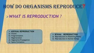
1. This principle was formulated in 1858 by German physician Rudolf Virchow.2. Cell reproduction is called cell division.3. Cell division has two major roles. It enables a fertilized egg to develop through various embryonic stages, and for an embryo to develop into an adult organism. It ensures the continuity from generation to generation; it is the basis of both asexual reproduction and sperm and egg formation in sexual reproduction.
8.3- Bacteria reproduce by binary fission.Bacterial chromosomes. Genes of bacteria are carried on one circular DNA molecule that is up to 500 times the length of the cell. Packaging is minimal: The DNA is complexed with a few proteins and attached to the plasma membrane at one point. Most of the DNA lies non-membrane bounded, in the center of the cell.
Binary fission. Prior to cell division, an exact copy of the chromosome is made. The attachment point divides so that the two new chromosomes are attached on adjacent parts of the plasma membrane. As the cell elongates and new plasma membrane is added, the attachment points of the two chromosomes move apart. Finally, the plasma membrane and new cell wall "pinch" through the cell, separating the two chromosomes into two new, genetically identical cells.
8.4- Eukaryotes have multiple chromosomes that are large and complex. Whereas a typical bacterium might have 3000 genes, human cells have 50,000100,000. These genes are organized into several separate, linear chromosomes that are found inside the nucleus. The DNA in eukaryotic chromosomes is complexed with protein in a much more complicated manner. This organizes and allows expression of much greater numbers of genes. The chromosomes are only visible under the light microscope just prior to and during cell division (Figure 8.4).
In multicellular plants and animals, the body cells (somatic cells) contain twice the number of chromosomes as the sex cells. Humans have 46 chromosomes in their somatic cells and 23 chromosomes in their sex cells. Different species may have different numbers of chromosomes.
8.5- Chromosomes duplicate and then split in two as a cell divides (Module 8.5).The DNA molecule in each chromosome is copied prior to the chromosomes' becomingvisible. As the chromosomes become visible, each is seen to be composed of two identical sister chromatids, attached at the centromere (Figure 8.5A). It is the sister chromatids that are parceled out to the daughter cells (the chromatids are thenreferred to as chromosomes). Each new cell gets a complete set of identical chromosomes.
8.6- The cell cycle multiplies cells. Most cells in growing, and fully grown, organisms divide on a regular basis (once an hour, once a day), although some have stopped dividing. Such dividing cells undergo a cycle, a sequence of steps that is repeated from the time of one division to the time of the next. Interphase represents 90% or more of the total cycle time and is divided into G1, S, and G2 subphases.
During G1, the cell increases its supply of proteins and organelles and grows in size.
During S, DNA synthesis (replication) occurs. During G2, the cell continues to prepare for the actual division, increasing the supply of other proteins, particularly those used in the process. Cell division itself is called the mitotic phase and involves two subprocesses, mitosis (nuclear division) and cytokinesis (cytoplasmic division). The overall result is two daughter cells, each with identical chromosomes. Mitosis is very accurate. In yeasts, one error occurs every 100,000 divisions.
8.7- Cell division is a continuum of dynamic changes. Following are the established dividing points for the phases of the cell cycle (mitosis includes prophase through telophase):
Interphase: duplication of the genetic material; ends when chromosomes begin to become visible.
Prophase: mitotic spindle is forming, emerging from microtubule organizing centers(MTOCs). Prophase ends when the chromatin has completely coiled into chromosomes;nucleoli are gone; nuclear membrane and nucleoli have dissolved.
Metaphase: spindle fully formed; chromosomes are aligned single file with centromereson the metaphase plate (the plane that cuts the spindle's equator). Anaphase: chromosome separation, from centromere dividing to arrival at poles. Telophase: the reverse of prophase.
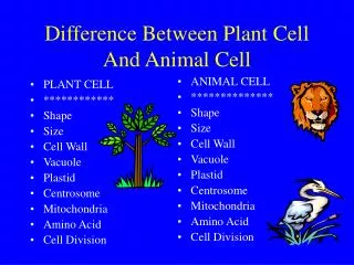
8.8- Cytokinesis differs for plant and animal cells. In animals, a ring of microfilaments contracts around the periphery of the cell, forming a cleavage furrow that eventually cleaves the cytoplasm.In plants, vesicles containing cell wall material collect among the spindle microtubules, in the center of the cell, then gradually fuse, from the inside out, forming a cell plate which gradually develops into a new wall between the two new cells. The membranes surrounding the vesicles fuse to form the new parts of the plasma membrane.
8.9- Anchorage, cell density, and chemical growth factors affect cell division.To grow and develop, or replenish and repair tissues, multicellular plants and animals must control when and where cell divisions take place. Most animal and plant cells will not divide unless they are in contact with a solid surface; this is known as anchorage dependence. Laboratory studies show that cells usually stop dividing when a single layer is formed and the cells touch each other. This density-dependent inhibition of cell growth is controlled by the depletion of growth factor proteins in masses of crowded cells.
8.10- Growth factors signal the cell-cycle control system. The cell-cycle control system regulates the events of the cell cycle. Three major checkpoints exist: (a) at G1 of interphase (b) at G2 of interphase (c) at the M phase. This regulation is a type of signal transduction. If, at these checkpoints, a growth factor is released, the cell cycle will continue. If a growth factor is not released, the cell cycle will stop, e.g. Nerve and muscle cells are non-dividing cells stuck at the G1 checkpoint.
8.11- Growing out of control, cancer cells produce malignant tumors. Cancer is a general term for many diseases in multicellular animals and plants involving uncontrolled cell division with the resultant tumor metastasizing. Cancer cells grown in culture are not affected by the growth factors that regulate density-dependent inhibition of cell division. A malignant tumor consists of cancerous cells. These tumors metastasize. This is in contrast to benign tumors, which do not metastasize.
When someone dies of cancer, they rarely die as a result of the primary tumor; it is usually the metastases that kills them. Cancers are named according to the tissue or organ of origin. Some cancer cells actually continually synthesize factors that keep them dividing.
Radiation and chemotherapy are two treatments for cancer. Radiation disrupts the process of cell division, and since cancer cells divide more often than most normal cells, they are more likely to be affected by radiation. Chemotherapy involves drugs that, like radiation, disrupt cell division. Some of these drugs for example, taxol target the mitotic spindle.
8.12- This is a review of the first part of the chapter. Read it for yourself.
Module 8.13 Chromosomes are matched in homologous pairs. Homologous chromosomes share shape, genetic loci, and carry genes controlling the same inherited characteristics. Each of the homologues is inherited from a separate parent. The sets are combined in the first cell following fertilization and passed down together from cell to cell during growth and development by mitosis.
In humans, 22 pairs, found in males and females, are autosomes. Two other chromosomes are sex chromosomes. in females, there are two X chromosomes in males, an X and a Y.
Module 8.14- Gametes have a single set of chromosomes. Adult animals have somatic cells with two sets of homologues (diploid, 2n). Sex cells (gametes = eggs and sperm) have one set of homologues (haploid, n). These cells are produced by meiosis. Sexual life cycles involve the alternation between a diploid phase and a haploid phase. The fusion of haploid gametes in the process of fertilization results in the formation of a diploid zygote.
Module 8.15- Meiosis reduces the chromosome number from diploid to haploid.Meiosis is a form of cell division that is used in sexual reproduction.Strategy: cell division twice - with reduction of chromosome number to 1/2.
Meiosis occurs only in diploid cells. Like mitosis, meiosis is preceded by a single duplication of the chromosomes. The overall result is four daughter cells, each with half the number of chromosomes. The process is dynamic but may stop at certain phases for long periods of time. The process includes two consecutive divisions (meiosis I and meiosis II). The halving of the chromosome number occurs in meiosis I. Sister chromatids separate in meiosis II.
8.17- Independent orientation of chromosomes in meiosis and random fertilization lead to varied offspring.Mechanism: During prophase I of meiosis, each homologue pairs up with its "other."When they separate at anaphase I, maternally and paternally inherited homologues move to one pole or the other independently of other pairs.
Consequences: Given n chromosomes, there are 2n ways that different combinations of the half-pairs can move to one pole. In humans, there are 223 (about 8.4 million) combinations of combining an individual's maternally inherited and paternally inherited homologues. Combining gametes into zygotes suggests there are 223 x 223 combinations in the zygote or about 64 trillion different zygotes that could result.
The consequences of the large amount of genetic variation generated by sexual reproduction is contrasted with the lower levels of genetic variation associated with asexual reproduction.
8.18- Homologous chromosomes carry different versions of genes.Example: coat color and eye color in mice. C (agouti = brown) and c (white) for different versions of the coat-color gene E (black) and e (pink) for different eye-color genes.In this example, with the information up to this point, there would be two possible outcomes for the genes on the two chromosomes in a gamete.
8.19- Crossing over further increases genetic variability. Crossing over is the exchange of corresponding segments between two homologues. The site of crossing over is called a chiasma. This happens between chromatids within tetrads as homologues pair up closely during synapsis (prophase I). Crossing over produces new combinations of genes (genetic recombination) (Figure 8.19B).