
LAB Exercises II-11 Digestive System Anatomy
E N D
Presentation Transcript
Biology 211 LabExercise II-11 Digestive System Anatomy
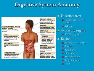
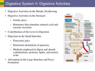
Introduction • The digestive system provides the body with the nutrients, water, and electrolytes essential for health • The organs of this system ingest, digest, and absorb food and eliminate the undigested remains as feces
What to know from the microscope: • Pancreas • acini(acinar cells) • islets of Langerhans • Salivary gland • stratified cuboidal epithelium • Liver • sinusoid • central vein, • hepatocytes • Stomach • gastric pit • simple columnar epithelium • smooth muscle • Ileum Slide • mucosa • submucosa • muscularisexterna ( circular and longitudinal smooth muscle layers) • serosa • villus(plural = villi)
Microscope Slides Pancreas Salivary gland Stratified cuboidal epithelium
Microscope Slides Liver Stomach Simple Columnar Epithelium
Microscope Slides Ileum Mucosa Submucosa Muscularisexterna Serosa villus
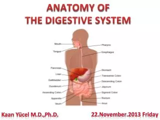
What to know from the model • Teeth • Dentin • Pulp • Enamel • Salivary glands • Submandibular • Sublingual, parotid • Tongue • Esophagus • Stomach • Rugae • Fundus • Body • Cardiac region • Pylorus • Greater and lesser curvatures • Longitudinal, circular, and oblique smooth muscle layer • Cardiac sphincter • Pyloric sphincter
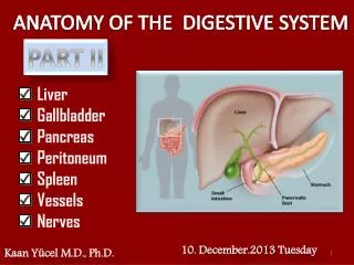
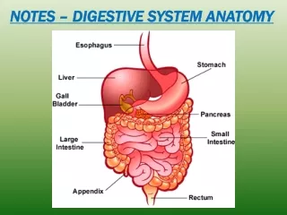
What to know from the model • Small intestine • Duodenum, jejunum, ileum • Plicaecirculares • Ileocecal valve • Large intestine • Cecum (appendix) • Ascending, transverse, descending, sigmoid colon • Haustra • Teniae coli • Rectum (anus) • Greater omentum • Mesentery • Liver • Left, right, caudate, quadrate lobes • Falciform ligaments • Hepatic portal vein • Left and right hepatic ducts • Common hepatic duct • Gallbladder • Cystic duct, common bile duct, hepatopancreatic duct • Pancreas • Head, tail, body, & pancreatic duct
Definitions • Teeth • Dentin - is a calcified tissue of the body • Pulp - made up of living soft tissue and cells called odontoblasts • Enamel - hardest and most highly mineralized substance of the body • Salivary glands • Submandibular - The salivary glands in mammals are exocrin glands, glands with ducts, that produce saliva. They also secrete amylase, an enzyme that breaks down starch into maltose • Sublingual • Parotid - largest of the salivary glands • Tongue • Esophagus - consists of a muscular tube through which food passes from the pharynx to the stomach
Definitions • Stomach • Rugae - refers to a series of ridges produced by folding of the wall of an organ • Fundus - is an anatomical term referring to the portion of an organ opposite from its opening • Body - anatomical region of the stomach in humans • Cardiac region - The opening of the esophagus into the stomach. • Pylorus - the region of the stomach that connects to the duodenum • Greater and lesser curvatures • Longitudinal, circular, and oblique smooth muscle layer • Cardiac sphincter - muscle fibers about the opening of the esophagus into the stomach • Pyloric sphincter - a sphincter at the opening from the stomach into the duodenum
Definitions • Small intestine - is the part of the gastrointestinal tract (gut) following the stomach and followed by the large intestine, and is where the vast majority of digestion and absorption of food takes place • Duodenum - first section of the small intestine • Jejunum - the middle section of the small intestine • Ileum - the final section of the small intestine • Plicaecircularesare- large flaps projecting into the lumen of the bowel. • Ileocecalvalveis-a sphincter muscle situated at the junction of the small intestine (ileum) and the large intestine. Its critical function is to limit the reflux of colonic contents into the ileum
Definitions • Large intestine • Cecum -is a pouch, connecting the ileum with the ascending colon of the large intestine • Ascending, transverse, descending, sigmoid colon • Haustra– the small pouches of the colon, which give it a segmented appearance • Teniaw coli - the three bands comprising the longitudinal layer of the tunica muscularis of the colon • Rectum (anus) • Greater omentum- is a large fold of peritoneum that hangs down from the stomach, and extends from the stomach to the posterior abdominal wall after associating with the transverse colon.
Definitions • Mesentery - is the double layer of peritoneum that suspends the jejunum and ileum from the posterior wall of the abdomen. • Liver • Left, right, caudate, quadrate lobes • Falciform ligaments - is a ligament which attaches the liver to the anterior body wall • Hepatic portal vein - is a vein in the abdominal cavity that drains blood from the gastrointestinal tract and spleen to the liver • Left and right hepatic ducts • Common hepatic duct - drains bile from the left functional lobe of the liver
Definitions • Gallbladder • Cystic duct- is the short duct that joins the gall bladder to the common bile duct • Common bile duct - a tube-like anatomic structure in the human gastrointestinal tract. It is formed by the union of the common hepatic duct and the cystic duct • Hepatopancreatic duct - is a duct joining the pancreas to the common bile duct to supply pancreatic juices which aid in digestion • Pancreas • Head, tail, body • Pancreatic duct - is a duct joining the pancreas to the common bile duct to supply pancreatic juices which aid in digestion
Hepatic portal vein Left hepatic duct Right hepatic duct Common hepatic duct Cystic duct
What to know from the pig • Teeth • Dentin • Pulp • Enamel • Salivary glands • Submandibular • Sublingual • Parotid • Tongue • Esophagus • Stomach • Rugae • Fundus • Body • Cardiac region • Pylorus • Greater and lesser curvatures • Cardiac sphincter • Pyloric sphincter
What to know from the pig • Small intestine • Duodenum, jejunum, ileum • Ileocecal valve • Large intestine • Cecum • Rectum (anus) • Greater omentum • Mesentery • Liver • Falciform ligaments • Gallbladder • Pancreas
The ileocecal valve is a sphincter muscle situated at the junction of the small intestine(ileum) and the large intestine Its critical function is to limit the reflux of colonic contents into the ileum I could not find a pig picture for this…but there is a really good picture for the cat on slide 50. It looks exactly the same for the pig
What to know from the cat • Tongue • Esophagus • Stomach • Greater and lesser curvatures • Cardiac sphincter • Pyloric sphincter • Small intestine • Duodenum, jejunum, ileum • Ileocecal valve • Large intestine • Cecum • Rectum (anus) • Greater omentum • Mesentery • Liver • Gallbladder • Pancreas