Download
1 / 22
220 likes | 501 Vues
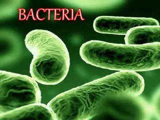
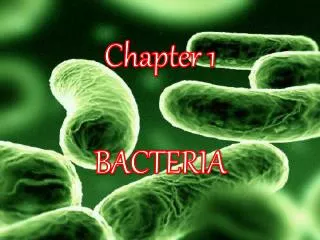
Structure and morphology of B acteria. Max Sanam. Perhaps the most elemental structural property of bacteria is cell morphology (shape). Typical examples include: coccus (spherical) bacillus ( rod-like) spirillum (spiral) filamentous.

E N D
Structure and morphology of Bacteria Max Sanam
Perhaps the most elemental structural property of bacteria is cell morphology (shape). • Typical examples include: • coccus (spherical) • bacillus (rod-like) • spirillum(spiral) • filamentous
Bacteria generally form distinctive cell morphologies when examined by light microscopy and distinct colony morphologies when grown on Petri plates. • These are often the first characteristics observed by a microbiologist to determine the identity of an unknown bacterial culture.
Cell wall • The cell envelope is composed of the plasma membrane and cell wall. • As in other organisms, the bacterial cell wall provides structural integrity to the cell. • In prokaryotes, the primary function of the cell wall is to protect the cell from internal turgor pressure caused by the much higher concentrations of proteins and other molecules inside the cell compared to its external environment.
The bacterial cell wall differs from that of all other organisms by the presence of peptidoglycan (poly-N-acetylglucosamine and N-acetylmuramic acid), which is located immediately outside of the cytoplasmic membrane. • Peptidoglycan is responsible for the rigidity of the bacterial cell wall and for the determination of cell shape.
Since the cell wall is required for bacterial survival, but is absent in eukaryotes, several antibiotics (penicillins and cephalosporins) stop bacterial infections • by interfering with cell wall synthesis, while having no effects on human cellsbecause human cells don't have cell walls, they only have cell membranes.
There are two main types of bacterial cell walls, Gram positive and Gram negative, which are differentiated by their Gram staining characteristics. • For both Gram-positive and Gram-negative bacteria, particles of approximately 2 nm can pass through the peptidoglycan • If the bacterial cell wall is entirely removed, it is called a protoplast while if it's partially removed, it is called a spheroplast. β-Lactam antibiotics such as penicillin inhibit the formation of peptidoglycan cross-links in the bacterial cell wall. • The enzyme lysozyme, found in human tears, also digests the cell wall of bacteria and is the body's main defense against eye infections.
The Gram positive cell wall • Gram positive cell walls are thick and peptidoglycans (mucopeptides, glycopeptides, mureins), the structural elements of almost all bacterial cell walls, constitute almost 95% of the cell wall in some Gram positive bacteria and as little as 5-10% of the cell wall in Gram negative bacteria. • Peptidoglycans are made up of a polysaccharide backbone consisting of alternating N-acetylmuramic acid(NAM) and N-acetylglucosamine(NAG) residues in equal amounts.
The cell wall of some Gram positive bacteria can be completely dissolved by lysozyme, as this enzyme attacks the bonds between GA and MA. • In other Gram positive bacteria, e.g. Staphylococcus aureus, the walls are resistant to the action of lysozyme. They have O-acetyl groups on carbon-6 of some MA residues.
The matrix substances in the walls of Gram positive bacteria may be polysaccharides or teichoic acids. • The latter are very widespread, but have been found only in Gram positive bacteria. There are two main types of teichoic acid: ribitolteichoic acids and glycerol teichoic acids. • The latter one is more widespread. • The exact function of teichoic acid is debated and not fully understood.
The Gram negative cell wall • Gram negative cell walls are thin and unlike the Gram positive cell walls, they contain a thin peptidoglycan layer adjacent to the cytoplasmic membrane. • This is responsible for the cell wall's inability to retain the crystal violet stain upon decolorisation (differentiation) with ethanol-acetic acid during Gram staining. • In addition to the peptidoglycan layer, the Gram negative cell wall also contains an outer membrane composed by phospholipids and lipopolysaccharides, which face into the external environment.
The chemical structure of the outer membrane lipopolysaccharides is often unique to specific bacterial strains and is responsible for many of the antigenic properties of these strains. • Gram negative cell walls consist of a few layers of peptidoglycan surrounded by a second lipid membrane containing lipopolysaccharides and lipoproteins. • Lipopolysaccharides, also called endotoxins, are composed of polysaccharides and lipid A (responsible for much of the toxicity of Gram-negative bacteria).
The plasma membrane or bacterial cytoplasmic membrane is composed of a phospholipidbilayer and thus has all of the general functions of a cell membrane such as acting as a permeability barrier for most molecules and serving as the location for the transport of molecules into the cell. • In addition to these functions, prokaryotic membranes also function in energy conservation • As a phospholipidbilayer, the lipid portion of the outer membrane is impermeable to charged molecules. However, channels called porins are present in the outer membrane that allow for passive transport of many ions, sugars and amino acids across the outer membrane.
Extracellular (external) structures Fimbrae and pili • Fimbrae (sometimes called "attachment pili") are protein tubes that extend out from the outer membrane in many members of the Proteobacteria. • They are generally short in length and present in high numbers about the entire bacterial cell surface. • Fimbraeusually function to facilitate the attachment of a bacterium to a surface (e.g. to form a biofilm) or to other cells (e.g. animal cells during pathogenesis) • Pili are involved in the process of bacterial conjugation where they are called conjugation pili or "sex pili".
Glycocalyx • Many bacteria secrete extracellular polymers outside of their cell walls called glycocalyx. • These polymers are usually composed of polysaccharides and sometimes protein. • Capsulesare relatively impermeable structures that cannot be stained with dyes such as India ink. • They are structures that help protect bacteria from phagocytosis and desiccation. • Slime layer is involved in attachment of bacteria to other cells or inanimate surfaces to form biofilms. Slime layers can also be used as a food reserve for the cell.
Flagella • Perhaps the most recognizable extracellular bacterial cell structures are flagella. Flagella are whip-like structures protruding from the bacterial cell wall and are responsible for bacterial motility • The arrangement of flagella about the bacterial cell is unique to the species observed. Common forms include: • Monotrichous - Single flagellum • Lophotrichous - A tuft of flagella found at one of the cell pole • Amphitrichous - Single flagellum found at each of two opposite poles • Peritrichous - Multiple flagella found at several locations about the cell
Intracellular (intenal) structures The bacterial chromosome and plasmids • Unlike eukaryotes, the bacterial chromosome is not enclosed inside of a membrane-bound nucleus but instead resides inside the bacterial cytoplasm. This means that the transfer of cellular information through the processes of translation, transcription and DNA replication all occur within the same compartment and can interact with other cytoplasmic structures, most notably ribosomes. • The bacterial chromosome is not packaged using histones to form chromatin as in eukaryotes but instead exists as a highly compact supercoiled structure, the precise nature of which remains unclear. • Most bacterial chromosomes are circular although some examples of linear chromosomes exist (e.g. Borreliaburgdorferi). • Along with chromosomal DNA, most bacteria also contain small independent pieces of DNA called plasmids that often encode for traits that are advantageous but not essential to their bacterial host. Plasmids can be easily gained or lost by a bacterium and can be transferred between bacteria as a form of horizontal gene transfer
Most bacterial chromosomes are circular although some examples of linear chromosomes exist (e.g. Borreliaburgdorferi). • Along with chromosomal DNA, most bacteria also contain small independent pieces of DNA called plasmids that often encode for traits that are advantageous but not essential to their bacterial host. • Plasmids can be easily gained or lost by a bacterium and can be transferred between bacteria as a form of horizontal gene transfer
Ribosomes and other multiprotein complexes • In most bacteria the most numerous intracellular structure is the ribosome, the site of protein synthesis in all living organisms. • All prokaryotes have 70S (where S=Svedberg units) ribosomes while eukaryotes contain larger 80S ribosomes in their cytosol. • The 70S ribosome is made up of a 50S and 30S subunits. • The 50S subunit contains the 23S and 5S rRNA while the 30S subunit contains the 16S rRNA. • These rRNA molecules differ in size in eukaryotes and are complexed with a large number of ribosomal proteins, the number and type of which can vary slightly between organisms. • While the ribosome is the most commonly observed intracellular multiprotein complex in bacteria other large complexes do occur and can sometimes be seen using microscopy.