Right leg re-vascularization
120 likes | 289 Vues
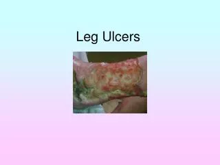
Right leg re-vascularization. 55 y-o male with 30yr history of IDDM and 10 years of ESRD with a twelve year old non-healing ulcer of the right foot with daily drainage requiring dressing changes.

Right leg re-vascularization
E N D
Presentation Transcript
Right leg re-vascularization 55 y-o male with 30yr history of IDDM and 10 years of ESRD with a twelve year old non-healing ulcer of the right foot with daily drainage requiring dressing changes. MRI revealed no evidence of osteomyelitis and the left groin was accessed for arteriography. After bilateral run-off RLE intervention was performed.
Via the Left groin the right leg run-off was accessed and diagnostic arteriogram revealed high-grade stenoses in the Right Superficial Femoral artery.
Subtracted images again revealing the Right SFA stenoses (blue). The subtraction artifact from the orthopedic screws are noted (black). The small curvy vessels are collateral vessels suggestive of even more distal disease (gray curved arrow).
At the level of the Right knee (screws are the white artifacts), the Right Popliteal artery is occluded (blue arrow) and only a late re-constitution of the Right Anterior Tibial (black arrow) artery is noted. These findings are consistent with the patient’s ischemic changes. There should be a wide-open Popliteal artery and three vessels running down the calf normally- the Right Anterior and Posterior Tibial arteries and the Peroneal artery. Only one “named” vessel is re-constituted pre-procedure.
After a long sheath was placed via the Left groin 5mm diffuse angioplasty was performed of the Right SFA followed by placement with a self-expanding nitinol stent. Follow-up arteriography demonstrates wide patency and continuous run-off.
The Right Popliteal artery (blue) was re-canalized using a sub-intimal technique with balloon 3.5mm angioplasty. As you can see, the Right Popliteal is now patent and continuous with the Right Anterior Tibial artery (white) and Tibio-Peroneal trunk (black).
S/P Right Superficial Femoral Artery PTA/Stent and Right Popliteal Artery re-canalization and PTA, there is now continuous run-off via the Right Anterior Tibial (black) and Right Posterior Tibial arteries (white).
Post Right leg re-vascularization This last picture is two weeks post re-vascularization. All drainage had ceased and the ulcer was healing nicely. After four weeks the ulcer had completely healed. The patient is maintained on Plavix 75mg daily. Blood Flow=Oxygen=Healing/ Tissue Preservation