Microorganisms
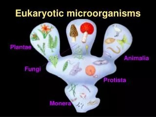
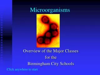
Microorganisms. Classification of Microorganisms: Microbes can be classified into four major groups: 1- Protozoa 2- Bacteria. 3- Fungi. 4- Viruses. 1- The Protozoa:

Microorganisms
E N D
Presentation Transcript
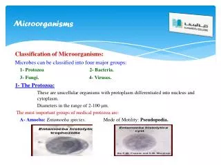
Microorganisms Classification of Microorganisms: Microbes can be classified into four major groups: 1- Protozoa 2- Bacteria. 3- Fungi. 4- Viruses. 1- The Protozoa: These are unicellular organisms with protoplasm differentiated into nucleus and cytoplasm. Diameters in the range of 2-100 μm. The most important groups of medical protozoa are: A-Amoeba:Entamoeba species. Mode ofMotility: Pseudopodia.
Protozoa B- Mastigophora: Mode of Motility: the Flagella • Gastrointestinal flagellates: Giardiaintestinalis • Urogenital flagellates: Trichomonasvaginalis • Tissue and blood flagellates: Trypanosoma, Leishmania
Protozoa Trypanosoma, andLeishmania.
Protozoa C- Ciliophora: Mode of Motility: the Cilia. • Example: Balantidium coli.
Protozoa D- Sporozoa: intracellular infection. Example: Plasmodiumthat cause Malaria.
Bacteria 2- The Bacteria: Bacteria are unicellular prokaryotic microorganisms that multiply by binary fission. Bacteria can be classified according to morphology, arrangement, and staining reaction into the following groups: 1- Filamentous bacteria:Streptomyces: antibiotic producers. 2- True bacteria: Cocci: Gram positive: Staphylococcus, Streptococcus. Gram negative: Neisseria. Bacilli: Gram positive: Bacillus, Clostridum, Corynebacterium. Gram negative: Enterobacteriaceae, Brucella. 3- Spirochetes:Slender flexuous spiral bacteria. Borrelia, Treponema, Leptospira. 4- Mycoplasma: The Smallest bacteria that lack of a rigid cell wall. 5- RickettsiaeandChlamydiae:intracellular parasites.
Fungi 3- The Fungi: These are saprophytic or parasitic organisms possessing relatively rigid cell walls. Medical fungi can be divided into: 1- Mould:Branching filaments; hyphae, mycelium. Usually 2 to 10 μm in width. Example: Epidermophyton, Trichophyton,Microsporum, Aspergillus. 2- True Yeasts: These are ovoid or spherical cells that reproduce asexually by budding and sexually with formation of spores. Example : Cryptococcus spp. 3- Dimorphic fungi: Produce a vegetative mycelium in artificial media, but are yeast like in infected lesions. Example: Histoplasma. 4- Yeast- like fungi: Example:Candida ( Pseudomycelium).
Viruses 4- The Viruses: Viruses consist of DNA or RNA enclosed in a simple protein shell known as a capsid. General properties of viruses: They are very small in size, from 20-300 m. They contain one kind of nucleic acid (RNA or DNA) as their genome. They are metabolically inert They are obligate intracellular parasites. They are only seen by electron microscope. Depend on the parasitized cell for survival and multiplication
Bacterial Morphology Structure of bacterial cells:Size, Shape, and Arrangement of bacterial cells: Morphology and arrangement of bacterial cells are criteria used for classification of bacteria into following groups: 1. Cocci (Singular: coccus). 2. Rods (bacilli), (Singular: rod, bacillus). 3. Vibrios (Singular: vibrio). 4. Spirilla (singular :Spirillum) 5. Spirochetes. (Singular: Spirochaete). 1. Cocci: These are round or oval bacteria measuring about 0.5-1.0 micrometer in diameter. When they multiplying, coccimay form pairs, chains, or irregular groups.
Bacterial Morphology Cocci in pairs are called diplococci, for example, meningococciand gonococci. Cocci in chains are called streptococci, for example Streptococcuspyogens. Cocciin irregular groups are called Staphytococci,for example, Staphylococcus aureus.
Bacterial Morphology 2. Rods (bacilli): These are stick-like bacteria with rounded, square, or swollen ends. They measure 1-10 micrometer in length by 0.3-1.0 micrometer in width. It may arranged in: Chains, for example, Streptobacillus species. Branching chains, for example, lactobacilli. Mass together, for example, Mycobacterium leprae. Remain attached at various angles resembling Chinese letters, for example, Corynebacterium diphtheria.
Bacterial Morphology 3-Vibrios: These are small slightly curved rods measuring 3-4 micrometer in length by 0.5 micrometers in width. Most vibrios are motile with a single flagellum at one end. They show a rapid darting motility. For example: Vibrio cholerae.
Bacterial Morphology 4-Spirochetes: These are flexible, coiled, motile organism, 6-20 micrometer in length. They progress by rapid body movements. Spirochetes are divided into three main groups: 1-Treponemes. 2- Borreliae. 3-Leptospires.
Bacterial structure The Bacterial Cell Envelope: This term refers to all materials and layers enclosing the cytoplasm. The most prominent of which are: 1- Cell wall. 2- Cell membrane. 1) Bacterial cell wall: It is the outer covering layer of the bacterial cell. It is a rigid structure consisting of two layers in Gram-positive bacteria and of three layers in Gram-negative. It is composed of a cross-linked polymeric mesh (Peptidoglycan) Peptidoglycan is long polymers of two sugar derivatives, NAG (N-Acetyl Glucosamine) and NAM (N-Acetyl Muramic acid) with side chains of four alternating D and L amino acids attached to NAM.
Bacterial Cell Wall The bacterial cell wall structure:
Bacterial Cell Wall Difference between Gram positive and Gram negative cell wall: The Gram-positive cell wall is formed of: Peptidoglycan layer: Exterior to cytoplasmic membrane. - Very thick in gram +vebacteria.- Constituting 50-60% of the cell wall. - Responsible for rigidity of the cell wall. Teichoicacid (somatic O antigen). The Gram-negative cell wall is formed of: Outer membrane of lipopolysaccharides (LPO): containing lipid A (endotoxin) & polysaccharide which represents somatic O antigen. Inner (cytoplasmic) membrane. The Periplasmicspace: separates outer and inner membranes. Peptidoglycan layer: - Located between the two membranes in the periplasmic space. - It is thin layer constituting 5-10% of the thickness of the cell wall.
Bacterial Cell Wall Difference between Gram positive and Gram negative cell wall: The Gram +ve cell wall The Gram –ve cell wall
Bacterial Cell Wall Functions of the bacterial cell wall: It maintains the shape of the bacterial cell. It protects the bacterial cell from external environmental hypotonicity because the cell wall is osmotically insensitive. It is responsible for gram stain reaction. It is responsible for endotoxic activity of gram negative bacteria. It plays a role in cell division.
Bacterial Cell Membrane • 2) Bacterial cell membrane:- It is a thin elastic semipermeable membrane that is internal to cell wall & surrounds the cytoplasm. • - It is consisted of phospholipids bilayers in which proteins (integral and peripheral protein) and enzymes are embeded. • The major functions of cytoplasmic • membrane are: • 1- Selective permeability to different molecules. • 2- Active transport of nutrients through special enzymes . • 3- Electron transport by its electron transport chain. • 4- Secretion & excretion of toxins and • hydrolytic enzymes outside the cell. • 5- It supplies the cell with energy, • It is the site of respiration. • 6- It provides enzymes needed for cell wall synthesis. • 7- It plays a role in DNA replication.
Glycocalyx and Capsule Structures external to the Cell Wall:( glycocalyx, flagella, axial filaments, and pili): 1- Glycocalyx and Capsule: It is a gelatinous viscous layer formed outside the cell wall of some bacteria. Many bacteria synthesize large amounts of extracellular polymers when growing in their natural environments. These polymers form capsules or glycocalyx. Its chemical nature may be: - - Polysaccharide as in the Pneumococcus, or - Polypeptide as in Bacillus anthracis. When these polymers closely surrounding the cell with well-organized structure; it is called Capsule, But if these polymers form a loose meshwork of fibrils extending outward from the cell and amorphous; it is called Glycocalyx.
Glycocalyx and Capsule Function of capsule or glycocalyx: • 1- Protection of bacterial cells from phagocytosis and antibodies. • 2- Diffusion barrier against some antibiotics. Capsule and glycocalyx can be demonstrated by light microscope using of capsule stain of bacterial smear or India ink wet mount. • In gram stained film, it appears as unstained halo around the organism.
Flagella 2- Flagella and motility: Motile bacteria possess filamentous appendages known as flagella, which act as organs of locomotion. The flagellum is a long, thin filament, twisted spirally in an open, regular waveform. Composed of several thousand molecules of the protein flagellin. It is about 0.02 μm thick and is usually several times the length of the bacterial cell. According to the species, there may be one, or up to 20, flagella per cell.
Flagella Flagella may be classified according to there arrangement as follow: 1- Monotrichous (single polar flagellum). 2- Lophotrichous (tuft of polar flagella). 3- Amphitrichous (one flagellum at each side of cell). 4- Peritrichous (flagella distributed over the cell). - Flagella are associated with chemotaxis process (chemical attraction)of bacterial cells that contribute in disease pathogenesis. - Flagella are highly antigenic.
Flagella Monotrichousflagellum Lophotrichous flagella Peritrichous flagella
Axial filaments 3- Axial filaments: • Some types of bacteria have a flagellum that lie inside periplasmic space • (over cell wall peptidoglycan and under the outer membrane). • This flagellum called endoflagellum or axial filament. • The endoflagella are more than half the length of the organisms and run along the axial aspect of the spiral body. • They are responsible for rotary motility(corck-screw like motility) of these organisms.
Pili and Fimbriae 4- Pili and Fimbriae: Many bacteria possess filamentous appendages termed pili or fimbriae. These are hair like projections that are shorter, thinner and more numerous than flagella (e.g. 100-500, being bornesurrounding each cell) originating from the cell surface & are found more in gram –ve bacteria. They are from 0.1 to 1.5 μm in length and having a uniform width between 4 and 8 nm. Fimbriae are of two types according to their functions: 1. Ordinary pili: which are important in mediating adhesion between the bacteria and host cells ( hemagglutination phenomenon). They are virulence factors of bacteria. 2. Sex pili: they are hallow, longer and thicker than ordinary pili. They initiate the process of conjugation ( genetic material exchange between bacteria).i.e. their function is to transfer DNA from F+ to F- bacteria.