Contrast media 1
Contrast media 1. Radiographic Contrast Media. RAD TECH 255 SPECIAL PROCEDURES MERRILLS VOL2 RTA BOOK CH 19. Subject Contrast. Range of differences in the intensity of the x-ray beam, after it has been attenuated by the subject (patient). Low Subject Contrast .

Contrast media 1
E N D
Presentation Transcript
Radiographic Contrast Media RAD TECH 255 SPECIAL PROCEDURES MERRILLS VOL2 RTA BOOK CH 19
Subject Contrast • Range of differences in the intensity of the x-ray beam, after it has been attenuated by the subject (patient).
Low Subject Contrast What can be done to attain medical information- • see the difference between muscle, organs or vessels Define and outline – organ structure and function
Contrast media • Defines subtle differences in subject contrast • Increases atomic number of area injected • Results in a SHORTER scale of subject contrast
Purpose of Contrast Media • To enhance subject contrast or render high subject contrast in a tissue that normally has low subject contrast.
Atomic Number • Fat = 6.46 • Water = 7.51 • Muscle = 7.64 • Bone = 12.31
Radiographic Contrast : Influenced by… • Radiation Quality (KVP) • Film Contrast • Radiographic object (Patient)
KVPTYPE OF CONTRAST USED DETERMINES KVP RANGE BARIUM 90 – 120 kVp IODINES 70 – 80 kVp (Ionic / Nonionic Water or Oil)
INJECTABLECONTRAST MEDIAfor RT 255 procedures INVASIVE PROCEDURES The “o-grams”
ALWAYS TAKE A “SCOUT” BEFORE CONTRAST INJECTION √ PATHOLOGY √TECHNIQUE √PREP & PRIOR CONTRAST √POSITIONING
SPECIAL “o-grams” • Venogram • Arthrogram • Sialogram • Myelogram • Arteriogram • Angiogram • Galactogram • Hystersalpingogram…….. etc
SPECIAL PROCEDURESARE INVASIVE ALWAYS GET PATIENT’S HISTORY AND CONSENT BEFORE BEGINNING OR GIVING ANY CONTRAST MEDIA
CONSENTS • SIGNED AND WITNESSED • AFTER PROCEDURE HAS BEEN EXPLAINED • CHECK DEPARTMENT PROTOCOL • WHO’S RESPONSIBLE ??????
CONSENTS • ASSAULT verbal threat of harm • BATTERY Unlawful touching - unauthorized treatment “X-RAY” TAKEN ON WRONG PATIENT • FALSE IMPRISONMENT Restraints require permission from patient or authorized person
BLOOD WORKLAB TESTS to check function of kidneys prior to injection of contrast • WATCH THE UPPER LIMITS • BUN = BLOOD UREA NITROGEN • Merrills pg 214 range is 8 to 25 pg 242 range is 10 - 20 always check with RAD when level above 20 • CREATININE levels range: • pg 214(0.6 - 1.5)pg 242(0.05 - 1.2) always check with RAD when level above 1.2 • Indicates function of kidneys • Diseases / dehydration / kidney failure
EGFR (new test) • Estimated • Glomerular • Filtration • Rate • More advanced test for • CREATININE levels
Review of Contrast Agents Types of Contrast Routes of Adminstration Chemical Components
Contrast Media changes the density of the organs Therefore changing the Subject contrast will change the Radiographic contrast and film contrast May need to INCREASE TECHNIQUE FROM SCOUT IMAGE
Negative contrast (AIR OR CO2) Radiolucent Low atomic # material Black on film Positive contrast (all others) Radiopaque High atomic # material White on film Contrast Media (review)
Radiolucent- negative contrast agent x-rays easily penetrate areas- appear dark on films Negative Contrast Media Air and gas complications emboli-air pockets in vessels lack of oxygen Radiopaque- positive contrast agent- absorbs x-rays appears light Positive Contrast Agents BARIUM IODINES Both + & - can be used in same study Types of Contrast Media
BARUIM Z# 56 NON WATER SOLUABLE GI TRACT ONLY INGESTED OR RECTALLY KVP 90 – 120* IODINE Z# 53 WATER SOLUABLE POWDER LIQUID INTRAVENOUS OR Intrathecal GI TRACT Also OIL based KVP BELOW 90* 2 BASIC TYPESOF CONTRAST material
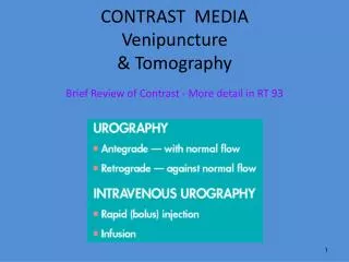
Methods of Administrationof Contrast Material • INGESTED • (ORAL) • RETROGRADE • AGAINST NORMAL FLOW • INTRATHECAL • Spinal canal • PARENTERAL (IV, Intrathecal) • Injecting into bloodstream • (anything other than oral)
Contrast media for SPECIAL PROCEDURES Diagnostic agents that are injected into • Circulatory System, Joint Spaces, Ducts • Body orifices/organs: uterus, breast, salivary & lymph glands
BARIUM – a review BARUIM SULFATE Not used in Special Procedures
Barium Sulfate • High atomic number • Not soluble in water • Used to coat the lining of organs • Supplied in different thicknesses • Used • Esophogram, UGI, Small Bowel,Lower GI or BE
Ba ADVERSE REACTIONS • BARIUM INERT • SUSPENSION MAY CAUSE ALLERGY • OCG TABLETS (IODINE) ALLERGY • AFTER EXAM – MAY SOLIDIFY DIFFICULT TO EVACUATE • INCREASE FLUIDS, MILD LAXATIVE • EXTRAVASATION OF CONTRAST INTO PERITONEUM
Ingested CONTRASTGastrografin or Hypaque • High atomic # • Close to iodine • Water soluble • Similar usage as Barium
GASTROGRAFINAdverse Reactions • Water soluble, safe in the abdominal cavity • Safe to use if perforation is suspected • Very harmful to the lung tissue • Do not use if aspiration is possible
Bowel Obstruction Note contrast Seen in kidneys as well Gastro – Pathology present
IODINEIONIC OR NON IONIC WATER OR OIL BASE
WATER BASED INJECTED VESSELLS/DUCTS INGESTED Organ function/flow OPEN WOUNDS OIL BASED INJECTED NEVER VESSELLS ONLY DUCTS NOT INGESTED OPEN WOUNDS IODINATED CONTRASTiodine z # 53
ALWAYS A WATER BASED IODINATED COMPOUND BOLUS INJECTION INFUSION DRIP IONIC VS NON IONIC CONTRAST 50 -70 % CONCENTRATE INJECTION OF IODINEinto Vessels
IONIC LESS $$$ MORE REACTIONS NON IONIC MORE $$$ LESS REACTIONS IODINE WATER BASED CONTRAST
CONTRAST MEDIAIODINE is either: IONIC or NON-IONIC • Osmolarity • # Of Particles (Cations + And Anions -) • In Solution Per Kilogram Of Water • High Osmolarity • =more Cations And Anions • Can Upset Homeostasis • Nonionic Have No Charged Particles
IONIC High Osmolality (Higher risk of complications) Diatrizoate sodium (Hypaque) Iothalamate meglumine (Conray) NON-IONIC Low Osmolality (Lower risk of complications) Gadodiamide (Omniscan) Iodixanol (Visipaque) Iopamidol (Isovue) Iopromide (Ultravist) Ioversol (Optiray) Contrast Agents
Less money More reactions More money Less reactions
OIL – BASED IODINECONTAST Instilled in ORGAN
Oil Based Iodine • Fatty Acids • Insoluble in water • White on the radiograph = Radiopaque • Uses • Broncography (lungs) • Tear ducts • Salivary glands • Lymphatic system • Hysterrosalpingogram • Galactography (breast ducts) • FAT EMBOLUS IF IT GETS INTO • BLOOD VESSEL