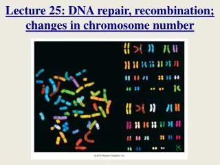
Changes in Chromosome Number
Changes in Chromosome Number. Chapter 3. Central Points. Chromosomes are composed of DNA and proteins Most humans have 46 chromosomes Possible to test fetal chromosome number Extra chromosomes affect fetus Problems with genetic testing can result in lawsuits.

Changes in Chromosome Number
E N D
Presentation Transcript
Changes in Chromosome Number Chapter 3
Central Points • Chromosomes are composed of DNA and proteins • Most humans have 46 chromosomes • Possible to test fetal chromosome number • Extra chromosomes affect fetus • Problems with genetic testing can result in lawsuits
Case A: Results Worry Pregnant Woman • Martha, age 41, is 18-weeks pregnant • Increased risk of chromosomal abnormalities • Amniocentesis recommended • Test results: • NoDown syndrome • Fetus is XYY (Jacobs syndrome)
3.1 Chromosomes • Thread-like structures in nucleus • Carry genetic information • Humans have 46 • Parts • Centromere • p arm • q arm • Telomeres
p arm Centromere q arm Fig. 3-1, p. 43
Animation: How Cells Reproduce (chromosome structure and organization)
3.2 Changes in Chromosome Number • Eggs and sperm are produced by meiosis • Begin with two copies of each chromosome (46) • Two divisions meiosis I and meiosis II • Homologous chromosome pairs separate • Produces haploid cells with one copy of each chromosome (23)
Before cells begin meiosis, the chromosomes duplicate. As meiosis begins, chromosomes coil and shorten, and become visible in the microscope. Each chromosome has a matching partner and the two chromosomes may exchange parts (cross over) during this stage, called prophase I. p. 44
The chromosome pairs line up along the middle of the cell, and spindle fibers attach to the centromere of each pair. This stage is called metaphase I. p. 44
Members of each homologous pair separate and move toward opposite sides of the cell. This stage is called anaphase I. p. 44
The chromosomes reach opposite poles of the cell, and the nuclei begin to re-form. This stage is called telophase I. The cytoplasm divides, and two cells are formed. These cells have half the number of chromosomes of the original cells and are called haploid cells. p. 44
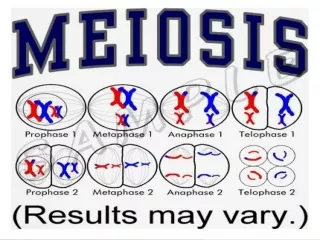
MEIOSIS I Before cells begin meiosis, the chromosomes duplicate. As meiosis begins, chromosomes coil and shorten, and become visible in the microscope. Each chromosome has a matching partner and the two chromosomes may exchange parts (cross over) during this stage, called prophase I. The chromosome pairs line up along the middle of the cell, and spindle fibers attach to the centromere of each pair. This stage is called metaphase I. Members of each homologous pair separate and move toward opposite sides of the cell. This stage is called anaphase I. The chromosomes reach opposite poles of the cell, and the nuclei begin to re-form. This stage is called telophase I. The cytoplasm divides, and two cells are formed. These cells have half the number of chromosomes of the original cells and are called haploid cells. Stepped Art p. 44
Two cells formed during meiosis I. In prophase II, the chromosomes of these cells become coiled, and move toward the center of the cell. p. 44
The 23 chromosomes in each cell attach to spindle fibers at their centromeres. This stage is called metaphase II. p. 44
Each centromere divides, and the newly formed chromosomes (also called sister chromatids) move to opposite ends of the cell. This stage is called anaphase II. p. 44
Finally, the chromosomes uncoil and the nuclear membrane re-forms. This stage is called telophase II. After the cytoplasm divides, the result is four cells, each with the haploid number of chromosomes. Meiosis is now completed. p. 44
MEIOSIS II Two cells formed during meiosis I. In prophase II, the chromosomes of these cells become coiled, and move toward the center of the cell. The 23 chromosomes in each cell attach to spindle fibers at their centromeres. This stage is called metaphase II. Each centromere divides, and the newly formed chromosomes (also called sister chromatids) move to opposite ends of the cell. This stage is called anaphase II. Finally, the chromosomes uncoil and the nuclear membrane re-forms. This stage is called telophase II. After the cytoplasm divides, the result is four cells, each with the haploid number of chromosomes. Meiosis is now completed. Stepped Art p. 44
Nondisjunction • Chromosomes fail to separate • Results in gametes and zygote with an abnormal chromosome number • Aneuploidy is variations in chromosome number that involve one or more chromosomes • Most aneuploidy from errors in meiosis
Chromosome number in gametes: Extra chromosome (n + 1) Extra chromosome (n + 1) Missing chromosome (n – 1) Missing chromosome (n – 1) Chromosomes align at metaphase I Nondisjunction at anaphase I Alignments at metaphase II Anaphase II Fig. 3-2, p. 45
Chromosome number in gametes: Extra chromosome (n + 1) Extra chromosome (n + 1) Missing chromosome (n – 1) Missing chromosome (n – 1) Chromosomes align at metaphase I Nondisjunction at anaphase I Alignments at metaphase II Anaphase II Stepped Art Fig. 3-2, p. 45
Aneuploidy • Effects vary by chromosomal condition • Many cause early miscarriages • Leading cause of mental retardation
3.3 ID of Chromosomal Abnormalities Two tests: • Amniocentesis (> 16 weeks) • Collects amniotic fluid • Fetal cells grown and karyotype produced • Chorionic villus sampling (CVS) (10–12 weeks) • Rapidly dividing cells • Karyotype within few days
Removal of about 20 ml of amniotic fluid containing suspended cells that were sloughed off from the fetus Biochemical analysis of the amniotic fluid after the fetal cells are separated out Centrifugation Fetal cells are removed from the solution Analysis of fetal cells to determine sex Cells are grown in an incubator Karyotype analysis p. 46
Removal of about 20 ml of amniotic fluid containing suspended cells that were sloughed off from the fetus Biochemical analysis of the amniotic fluid after the fetal cells are separated out Centrifugation Fetal cells are removed from the solution Analysis of fetal cells to determine sex Cells are grown in an incubator Karyotype analysis Stepped Art p. 46
Animation: Chromosomes and Human Inheritance (karyotype preparation)
Chorionic villi Ultrasound to monitor procedure Developing placenta Developing fetus Bladder Uterus Chorion Catheter Amniotic cavity Rectum p. 47
Amniocentesis Only Used in Certain Conditions • Risks for miscarriage; typically only done under one of following circumstances: • Mother > 35 • History of child with chromosomal abnormalities • Parent has abnormal chromosomes • Mother carries a X-linked disorder • History of infertility or multiple miscarriages
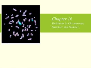
Other Chromosomal Variations • Polyploidy: multiple sets of chromosomes • Euploid: normal two copies of each chromosome • Trisomy: three copies of one chromosome • Monosomy: only one copy of a chromosome • Structural changes: duplication, deletion, inversion, translocation
Normal chromosome One segment repeated three times p. 47
Segment C deleted p. 47
Chromosome A Chromosome B Translocation p. 47
Animation: Meiosis and Sexual Reproduction (Meiosis I and II)
3.4 Effects of Changes in Chromosomes • Vary by chromosome and type of variation • May cause birth defects or fetal death • Monosomy of any autosome is fatal • Only a few trisomies result in live births