GASTROESOPHAGEAL REFLUX DISEASE. .
400 likes | 955 Vues
GASTROESOPHAGEAL REFLUX DISEASE. Definitions of Reflux. Clinical manifestations of reflux of stomach & duodenal contents into the esophagus. Characterized by any combination of symptoms, radiologic, endoscopic, or pathologic changes. In its milder forms, is common .

GASTROESOPHAGEAL REFLUX DISEASE. .
E N D
Presentation Transcript
Definitions of Reflux • Clinical manifestations of reflux of stomach & duodenal contents into the esophagus. • Characterized by any combination of symptoms, radiologic, endoscopic, or pathologic changes. • In its milder forms, is common. • Its most severe forms is uncommon but life-threatening. • GERD is preferable to "reflux esophagitis“ • GERD may be associated with a sliding HH, but "symptomatic HH" is an anatomic entity & not the underlying pathophysiology in GERD.
PATHOGENESIS. • The most common event : • Transient relaxation of the LES unassociated with swallowing or the distention of the esophagus. • 2 abnormalities: • A LES with very low tone & pressure. • Inappropriate relaxation of a normally competent sphincter. • Acid within the esophagus is cleared less well by patients with GERD than by normal subjects, although the manometric tracings in both groups seem identical.
PATHOGENESIS. • Gastric acid - pepsin important. • Bile salts & pancreatic enzymes, may be responsible if acid is absent. • The combination of bile salts plus acid is more injurious to the esophagus than either agent alone. • Other: • Altered or abnormal esophageal mucus • Abnormal saliva content. • Diminished resistance of the esophageal mucosa to digestion.
PATHOGENESIS: • Pregnancy: • From increased abdominal pressure by the fetus. • Diminished LES strength caused by increased estrogen & progesterone. • Weight gain also aggravate reflux through an unknown mechanism. • Resection of the lower esophageal area for cancer or myotomy for achalasia. • It is especially severe in progressive systemic sclerosis.
PATHOGENESIS. • Although HH may be associated with reflux, its presence is much less important, as it is present in a large percentage of normal subjects. • It is not necessary to spend time to find a HH with most patients with GERD. • Focus should be on the symptoms of reflux.
SYMPTOMS. • 1.Heartburn, the most common. • Vary from mild burning to chronic, severe markedly limiting a patient's lifestyle. • 2. Regurgitation of gastric contents, either into the mouth or into the respiratory tree: nocturnal wheezing, coughing, hoarseness, a need to clear the throat repeatedly, or a sensation of deep pressure at the base of the neck.
SYMPTOMS. • 3.Dysphagia is often present. • When severe, may indicate stricture,but even if mild & must be carefully sought. • Dysphagia is for solids, usually overcome by swallowing repeatedly or by washing the bolus down with water. • Many are aware of the location of each solid as it travels down the esophagus.
SYMPTOMS. • 4.Blood loss may result from esophageal erosions & shallow ulcers. • Rarely life-threatening hemorrhage & much likelychronic low grade, producing IDA. • Some have very few other clinical manifestations & discovered by endoscopy during evaluation of occult GIB. • Alcoholabuse produce severe erosive esophagitis with bleeding. • In these abstinence from alcohol is important.
DIAGNOSIS. • History &clinical manifestations are the most important . • Objective testing quantify the extent & severity. • In the majority, diagnosed by typical symptoms & the response to therapy. • Diagnostic evaluation becomes important when symptoms are atypical &/or do not respond to therapy. • Diagnosis include: • 1. Documenting reflux. • 2.Linking reflux to symptoms. • 3. Assessing the effect of reflux on eso mucosa.
1.DOCUMENTING REFLUX: Ba • Reflux during a barium swallow in adults is uncommon unless vigorous provocative maneuvers are employed. • When spontaneous reflux of barium is seen, it usually means free reflux. • The absence of reflux radiographically does not exclude GERD.
DOCUMENTING REFLUX. • The 24-hour monitoring of esophageal pH. • Relationship between symptoms (heartburn, chest pain, wheezing) & episodes of acid reflux confirmed. • Repeated - prolonged bursts of acid exposure suggest that abnormal GERD.
DOCUMENTING REFLUX. • In children - infants, reflux can be measured non invasively by RA 99mTc sulfur with or without augmentation by an abdominal binder if free reflux is not seen.
2.LINKING REFLUX TO SYMPTOMS. • If pain is the predominant symptom, rather than heartburn, a Bernstein acid infusion test may be performed.
3.ASSESSING THE EFFECT OF REFLUX ON THE ESOPHAGEAL MUCOSA. • A barium swallow detects gross changes, as stricture or ulcer, but misses shallow ulcerations - erosions, detected by OGD. • On OGD only lesions such as erosions & ulcerations should be taken as proof of esophageal damage, as erythema, edema, or friability, are subject to wide interobserver variation. • In 50% with moderate - severe symptoms, the mucosa appears absolutely normal, but a biopsy may demonstrate histologic changes(NERD).
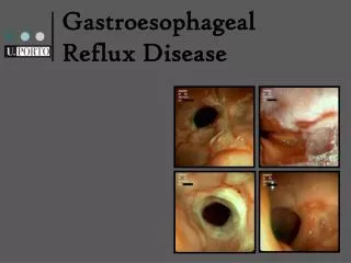
The LA Classification system– Grade A reflux esophagitis Grade A: One (or more) mucosal break, no longer than 5 mm, that does not extend between the tops of twomucosal folds. Stomach
The LA Classification system– Grade B reflux esophagitis GradeB: One (or more) mucosal break, more than 5 mm long, that does not extend between the tops of two mucosal folds. Stomach
The LA Classification system– Grade C reflux esophagitis Grade C: One (or more) mucosal break that is continuous betweenthe tops of two or more mucosal folds, but which involvesless than 75% of the circumference. Stomach
The LA Classification system– Grade D reflux esophagitis Grade D: One (or more) mucosal break that involves at least75% of the esophageal circumference. Stomach
APPROACH TO THE PATIENT • Endoscopy indicated if: • Hematemesis is present. • Prolonged & not respond to empiric treatment. • Systemic manifestations, as weight loss, anemia. • Occult blood–positive stool are present. • If the appearance of the esophageal mucosa is normal during endoscopy, biopsies can also be obtained to search for objective evidence of microscopic esophagitis (NERD).
APPROACH TO THE PATIENT • After first evaluation, it may be appropriate to begin empiric therapy • If the response to therapy is poor, esophageal pH monitoring can confirm the diagnosis. • At the same time, esophageal manometry may be performed to estimate LES pressure & to determine the presence or absence of peristaltic waves. • If dysphagia is present, a barium swallow is appropriate, Uncommonly, reflux, stricture or a deep ulcer seen, which leads to immediate endoscopy for more complete evaluation.
1.ESOPHAGEAL STRICTURE. • Only a few develop strictures. • Usually at the lower end, but sometimes migrating over years to the mid or higher. • Cause is Circumferential ulceration. • If reflux can be controlled, these strictures may disappear. • Dysphagia is the clinical hallmark. • The dysphagia tends to be constant ,slowly progressive, causing the patient to alter the type of food taken.
ESOPHAGEAL STRICTURE. • Most easily evaluated by barium swallow. • Sometimes the extent of the strictured area is overestimated unless the esophagus below the stricture can be fully distended by barium. • For mild strictures, the ingestion of barium-soaked bread or a bolus can draw attention to slight luminal narrowing where the bolus is impacted. • Endoscopy with biopsy &/or brush cytology is required to make certain that the stricture is benign.
2.ESOPHAGEAL ULCER. • The presence of an ulcer can be suspected on a barium swallow & confirmed endoscopically. • Characteristically produce severe & unrelenting pain, often with radiation of the pain to the back. • Brisk hemorrhage may be caused by erosion of an esophageal artery. • The ulcer usually is in columnar (Barrett's) epithelium.
3.BARRETT'S ESOPHAGUS (COLUMNAR EPITHELIUM). • The presence on biopsy of specialized columnar epithelium with goblet cells in the esophagus. • In some patients with chronic reflux esophagitis, the healing epithelium replaced with a specialized columnar epithelium with intestinal metaplasia. • The junctional zone between squamous & columnar (Barrett's) epithelium can progress upwards over years.
BARRETT'S ESOPHAGUS (COLUMNAR EPITHELIUM). • Identified endoscopically as salmon-pink (gastric-appearing) mucosa above the lower esophageal sphincter. • Barrett's epithelium is often found at & below mid-esophageal strictures & around deep esophageal ulcers. • Barrett's epithelium is a marker for severe reflux & a precursor to adenocarcinoma of the esophagus.
4.PULMONARY ASPIRATION. • Into the larynx & tracheobronchial tree. • Produces mild laryngeal or respiratory symptoms or hoarseness or intense respiratory stridor. • The gastric contents do not have to reach the larynx,as acid in the esophagus can cause closure of small bronchi by a vagal reflex. • Or volatile HCL can reach upper airways. • Wheezing, hoarseness, or coughing occur. • Dual esophageal pH monitoring with pH probes in both the lower & upper esophagus can help. • Treatment of reflux followed by disappearance of pulmonary symptoms may confirm the relationship.
Possible extraesophageal manifestations of GERD • Asthma • Sinusitis • Dental erosions • Reflux laryngitis • Vocal cord ulcers • Subglottal/tracheal stenosis • Laryngospasm Jailwala & Shaker 2000; Richter 2000; Ulualp et al 1999
Symptoms of Reflux in Infants • Regurgitation – emesis &weight loss • Esophagitis - chest pain, irritability, feeding problems, anemia, hematemesis, stricture causing obstruction • Neurobehavioral - infant “spells” (seizure-like events), Sandifer syndrome (opisthotonos & other abnormal posturing) • Respiratory symptoms - chronic or recurrent pneumonia, wheezing (especially intractable asthma), apnea (especially obstructive), cyanotic episodes, stridor, cough, hiccups, hoarseness • Complex respiratory disease-reflux interactions - esophageal atresia, TEF, cystic fibrosis.
TREATMENT OF GASTROESOPHAGEAL REFLUX DISEASE Step 1. Simple measures (lifestyle changes & nonsystemic treatment) A. Elevate head of bed B. Avoid food and fluid intake before bedtime C. Avoid cigarettes, coffee, alcohol D. Avoid chocolate, peppermint E. Avoid tight clothing around the waist F. Take antacids 1 hour after meals, at bedtime, and as needed G. Reduce fat in diet H. Lose weight Step 2. Measures for resistant cases (systemic treatment) Step 2a. H2-receptor antagonists A. Cimetidine, 300 mg q.i.d.* B. Ranitidine, 150 mg b.i.d.* C. Famotidine, 20 mg b.i.d.* D. Nizatidine, 150 mg b.i.d.* Step 2b. Prokinetic agents A. Metoclopramide, 10 mg q.i.d.* B. Cisapride, 10 mg q.i.d.* C. Bethanechol, 10 mg q.i.d.* Step 3. Measures for patients with GERD resistant to H2-receptor antagonists A. Proton pump inhibitor: omeprazole, 20 mg/day, or lansoprazole, 30 mg/day* Step 4. Measures for patients with GERD resistant to steps 1, 2, & 3 or patients who need long-term maintenance treatment A. Surgical fundoplication B.Endoscopic treatme
Endoscpic management of GERD: Endoscopic Baloon dilatation of esophageal stricture. Endoscopic photodynamic therapy, laser, or multipolar electrocoagulation ablasion of Barret esophagus. Endoscopic Radiofrequency application to LES. Laproscopic funduplication. Endoscopic antireflux stents.
Endoscopic therapies – the Stretta procedure Step 1 Step 2 Step 3
Severe postglottic edema Severe lingual tonsil hypertrophy Tracheal cobblestoning Arytenoid edema carinal blunting
Heartburn disturbs my sleep I cannot eat and drink whatever I like My whole life is affected GI symptoms bother me! I’m worriedand concerned I cannot bendover or exercise Illustrator: Eric Werner