Download
1 / 52
600 likes | 999 Vues
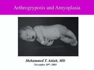
NEW DEVELOPMENTS IN ARTHROGRYPOSIS (MULTIPLE CONGENITAL CONTRACTURES). Judith G. Hall, OC, MD The University of British Columbia and BC Children’s Hospital Vancouver, BC Canada NO KNOWN CONFLICTS OF INTEREST. ARTHROGRYPOSIS (MULTIPLE CONGENITAL CONTRACTURES).

E N D
NEW DEVELOPMENTS IN ARTHROGRYPOSIS(MULTIPLE CONGENITAL CONTRACTURES) Judith G. Hall, OC, MD The University of British Columbia and BC Children’s Hospital Vancouver, BC Canada NO KNOWN CONFLICTS OF INTEREST
ARTHROGRYPOSIS(MULTIPLE CONGENITAL CONTRACTURES) Congenital nonprogressive limitation of movement of two or more joints in different body areas (lots and lots of things get included)
PLAN OF TALK • Frequency of congenital contractures • Ways to approach a diagnosis • Areas of body involved • Etiologic groupings • Similarity groupings • Genes – pathways/networks • Fetal akinesia deformation sequence • Fetal movement • Prenatal diagnosis and therapy • Effects of non-movement • Challenges NO KNOWN CONFLICTS OF INTEREST
CONGENITAL CONTRACTURES IN THE NEWBORN • Clubfoot…………………………….........1/500 – 1/1000 • Congenital dislocated hips....................1/200 – 1/500 • Multiple congenital contractures…..1/3000 – 1/6000 • All congenital contractures……………..1/100– 1/250
Arthrogryposis is not a diagnosis —it is a sign
FREQUENCY OF ARTHROGRYPOSIS- Many lethal types, miscarriages, stillborns • VERY heterogeneous • No proper ICD code(s) • Need population base • Australia epidemic • North America • Washington State • British Columbia registry • Others • Finland • Sweden
EPIDEMIOLOGY • Frequency ~ 1/3000 births • Prevalence ~ 1/4,000 – 6,000 • Geographic • Gender • Parental Age • “Outbreaks”
OCCURRENCE OF ARTHROGRYPOSIS • ~ 1/3000 – 5000….live births • 1/3…………………Amyoplasia • 1/3…………………CNS – newborn lethal • 1-3…………………Heterogeneous group of disorders
ARTHROGRYPOSIS STUDY GROUP 350 Study Group 500 Insufficient data 800 Secondary arthrogryposis >1500 cases Not congenital contractures From: Shriner’s Hospitals, Portland Spokane Children’s Orthopedic Hospital, Seattle University of Washington Hospital Artrhogryposis Association Correspondence, 1975-1978, University of Washington
Ways to approach a diagnosis – What are useful clinical discriminators?
APPROACH TO MULTIPLE CONGENITAL CONTRACTURES - CLINICAL • Mainly limbs • Limbs and other body areas • Limbs and CNS/lethal
AREAS OF INVOLVEMENT TOTAL STUDY GROUP 33% 33% 33%
AMYOPLASIA“CLASSICAL ARTHROGRYPOSIS” • Typical symmetric positions of limbs • Usually “teratogenic” clubfoot • Absent muscles with fibrotic replacement • Mid facial hemangioma • 10% abdominal structural anomaly (vascular accident) and other vascular compromise (lost fingers or toes) • Apparent increase in one of monozygotic twins • Surprisingly good response to early physical therapy • No apparent recurrence risk or risk for other congenital anomalies
WAYS TO APPROACH ARTHROGRYPOSIS BY ETIOLOGIC GROUPING • Muscle • Tendon length & placement • Peripheral nerve and end plate • CNS function • Bone • Limiting space • Maternal illness, medications or trauma • Vascular disruption
ARTHORGRYPOSIS ETIOLOGIC GROUPING - 1 • Muscle (myopathies, and distal arthrogryposes – TNNT3, TPM2, TNN12, MYH3 fast twitch muscle) • Tendon length & placement (Trismus pseudocamptodactyly) – MYH8 • Peripheral nerve and end plate (Multiple Pterygium Syndrome – Escobar type and lethal) (CHRNG, CHRNA1, CHRNBi, CHRND, RAPSN and antibodies to these) – compromises of Ach receptor • CNS function (Trisomy 18) • Limiting space (Assymetric) • Vascular compromise (Amyoplasia)
TYPES OF MUSCLE • Striated/voluntary • Smooth • Cardiac • Mixtures
ARTHORGRYPOSIS ETIOLOGIC GROUPING - 2 • Spinal cord – X-linked lethal – ubiquitin • Fetal akinesia deformation sequence • LCCS1 – 3, GLE1 (mRNA xport mediation), ERBB3, PIPSKIC (phosphotidyl inosotol pathway—also involved in muscle mRMA export) • Bone dysplasia (Diastrophic dysplasia) • Limiting space (Assymetric) • Maternal illness • Medication • Trauma
ARTHROGRYPOSIS – OBVIOUS OTHER WAYS OF GROUPING • Amyoplasia and other vascular compromise • Distal arthrogryposes • Pterygium syndromes • Bony fusions • Myopathies • Lethal SMA, X-linked lethal arthrogryposis • Lethal Pena Shokeir Phenotype (CNS structure subtypes) COFS (Cerebro Oculo Facial syndrome) LCCS 1-3 (Lethal Congenital Contracture syndromes) Neu Laxova syndrome • Camptodactylies • Skeletal dysplasias • Malformations syndromes • Chromosomal abnormalities
CLASSIFICATION OF DISTAL AMCs Hall Bamshad Gene I Distal 1A TPM2 IIA Gordon (cleft palate, SS) 3 IIB Ophthalmoplegia (fine muscle) 5 IIC Cleft Lip (10) IID Scoliosis + DA 4 IIE Trismus + Unusual Hand + DA 7B Freeman-Sheldon Syndome 2 MYH3 Sheldon-Hall 2B TNNT3, TNN12 Sheldon-Hall Look Alike 2C MYH3 Deafness + DA 6 11q25 Trismus Pseudocamptodactyly 7A MYH8 AD, Multiple Pterygium 8 Contractural Arachnodactyly 9 FBN2 Absent Teeth + DA (11) Chitayat, AR, DD (12) X-linked (13) Stavit, S. African, Naguib (14) Moore-Weaver Distal (15) MR, DD (16)
PTERYGIUM SYNDROMES TYPE INHERITANCE DISTINGUISHING GENE FEATURES Popliteal pterygium AD Clefts, lip pits normal nail IRF6 Antecubital pterygium AD Only elbows involved -- Mutiple pterygium (Escobar type) AR Cervical vertebral anomalies, CHRNG hands involved, chin- sternum pterygium, facies Lethal mutliple AR Extensive contractures, hypertelorism, pterygium CHRNG, chin-sternum pterygium, CHRNA1, small chest CHRNB1, RAPSN Lethal popliteal pterygium AR Facial cleft, syndactyly IRF6 (Bartoscas Papas) (hands and feet), genital anomaly Pterygium and ectodermal AR Fine sparse hair, nail anomalies -- dysplasia (hands and feet) Pterygium and malignant hyperthermia AR Torticolis, scoliosis, MH ?RYR1
FETAL AKINESIA DEFORMATION SEQUENCEPENA SHOKIER PHENOTYPE • Intrauterine growth retardation • Congenital contractures of the limbs • Hypoplastic lungs • Short umbilical cord • Polyhydramnios – short gut • Craniofacial anomalies • Micrognathia +/- small mouth • +/- cleft palate • High bridge of nose • Depressed tip of nose
Lack of normal mechanical forces may lead to secondary deformations • “Use” is essential for normal development
Maternal Connective tissue illness skeletal dysplasia Fetal Vascular crowding compromise Neurologic Muscle deficits defects LIMITATION OF FETAL JOINT MOBILITY MULTIPLE CONGENITAL CONTRACTURES (ARTHROGRYPOSIS)
HUMAN FETAL LIMB MOVEMENT • Starts 8 weeks, proximal limbs 9 weeks, distal 10 weeks • Requires intact neuromuscular unit • Maternal injury and CVS/early amniocentesis allow timing of limb involvement
EMBRYONIC LIMB DEVELOPMENT • Cranial caudal progression • Upper limbs before lower • Right side before left • Vascular supply to CNS shifting
EMBRYONIC/FETAL MOVEMENT Week 4 Heart beating begins 5-6 Head and trunk “stirs” 7 Shoulders “shrug” 8 Rhythmic “breathing” begins even though larynx not open Jaw starts to move 9 Upper arms moving 10 Hips, lower arms moving 11 Lower limbs kicking 12 Hands open and ankles moving into correct position
PRENATAL DIAGNOSIS BY ULTRASOUND(WHAT ARE THE CLUES, WHAT TO LOOK FOR) • Usually not picked up without long careful real time US study – 45 min – 1 hr • Nuchal edema • Thin undercalcified bones • Movement may start any time from 11 weeks to 34 weeks • Small lungs • Diaphragm defect or decreased movements • Other structure or space constraints (amniotic bands, uterine fibroid, amount of amniotic fluid)
As organs begin to function muscles begin to contract stretching developing tissues from inside and outside
DELIVERIES AT 39 WEEKS • Cephalic 60% • Breech 37% • Transverse 3% • C-section 45% • Fractures 10% - 20% • Birth weight 30th centile
Is intrauterine therapy possible? - “Physical Therapy in utero”-
DEPENDS ON • Functional CNS • Intact end plate • Functional muscles • Space to move
IN UTERO THERAPY • Fetal movement relates to maternal movement - Exercise - Deep breathing - Caffeine • Early delivery if lungs mature
FETAL MOVEMENT • Essential for normal development of limbs • Mechanical transduction of cells • Lack of movement leads to fetal akinesia deformation sequence • Maternal activity may affect outcome • Grace period of 3 – 4 months
CATCH-UP(3 – 4 MONTH WINDOW AFTER BIRTH) • Lung - avelolar growth • Gut - motility and absorption • Joints - loosening of contractures • Muscle - use reverses atrophy • Growth - bone mineralization, increase in length
LACK OF MECHANICAL FORCES LEADS TO DEFORMATION • “Use” is essential for normal development • Disuse leads to • Muscle atrophy • Increased connective tissue • Abnormal non-functional positions • Changes in joint surface
SECONDARY EFFECTS FROM LACK OF MOVEMENT IN UTERO • IUGR – limbs are short • Contractures with “collagenosis”, extra connective tissue, thick capsule • Abnormal relationship of limb to weight bearing – joints at odd angles • Muscle – disuse atrophy, decreased mass • Dimples – attached to overlying skin • Other changes of FADS – lungs, gut, craniofacial, etc.
EFFECTS OF FADS ON • On musculoskeletal system • On lungs • On gut • On growth • On development of motor skills
GROWTH • AMC affected limbs - short and small • Final height ~ 5th centile for family • Less muscle and less calcification of bone means less weight • Avoid obesity – makes for more work • Some limbs grow even less normally (like post-polio)
CHALLENGES – • Not miss opportunities • Stretching, weight bearing • Avoid muscle atrophy, night splints • Prevention • I°, II°, III° • Prenatal therapy • Avoid scarring • Not harm joint cartilage • Not allow atrophy of what is there • Keep from returning to “in utero position” • Intercede along mechanistic pathways • Cytokines, alternative metabolic pathways, fetal effects • Multi-system considerations • Enormous heterogeneity • But commonalities as well
FAMILY’S JOB • Ask questions • Take photographs • Make a notebook • Keep records • Ask questions
DOCUMENTATION • Photographs and videos • Notebook • Changes over time • New observations
If and only if a specific diagnosis cannot be made should a 5% recurrence risk estimation be given
Prognosis depends on the specific diagnosis and the natural history of that disorder
WEB ADDRESS FOR THE BOOK http://www.global-help.org/publications/ books/help_arthrogryposis.pdf
LAY GROUPS AROUND THE WORLD • Australia: http://www.taag.org.au/ • Germany, Austria & Switzerland: http://www.arthrogryposis.de/iga/info_en • Sweden: http://www.amcforeningen.se/ • UK: http://www.tagonline.org.uk/index.html
REFERENCES • Staheli LT, Hall JG, Jaffe KM, Paholke DO. Arthrogryposis: A text atlas. Cambridge University Press; Cambridge, UK, 1998. • Hall, JG. Arthrogryposes (Multiple congenital contractures). In: Emery and Rimoin’s principle and practice of medical genetics. Vol 3, 5th edition. Eds. Rimoin, DL, Connor JM, Pyeritz RE, Kork BR. Churchill Livingstone: New York, Chapter 168, p. 3785-3856, 2007. • Hall JG, Vincent A. Arthrogryposis. In: Neuromuscular diseases of infancy, childhood, adolescence – a clinician’s approach. Eds H Jones, DC De Vivo, BT Darris. Butterworth: Boston, Chapter 7, p. 123 – 141, 2003. • Hall JG. Arthrogryposis. In: Management of genetic syndromes, 2nd ed. Eds. Cassidy SB, Allanson JE. Wiley- Liss: Hoboken, NJ, Chapter 7, p. 63 – 86, 2005.