Human Genetics: Chromosomal Abnormalities and Cell Cycle Overview
280 likes | 336 Vues
Delve into human genetics to grasp chromosomal disorders like Klinefelter, Turner, and Down syndromes. Explore cell cycle stages such as Mitosis and Meiosis, and understand non-disjunction consequences. Learn about chromosomal abnormalities, monosomy, trisomy, and numerical sex chromosome disorders like Down, Turner, and Klinefelter syndromes. Discover the role of chromosomes in heredity and genetic material transfer. Watch informative videos on cell division and grasp the differences between Mitosis and Meiosis. Gain insights into non-disjunction and its impact on chromosome separation during meiosis.

Human Genetics: Chromosomal Abnormalities and Cell Cycle Overview
E N D
Presentation Transcript
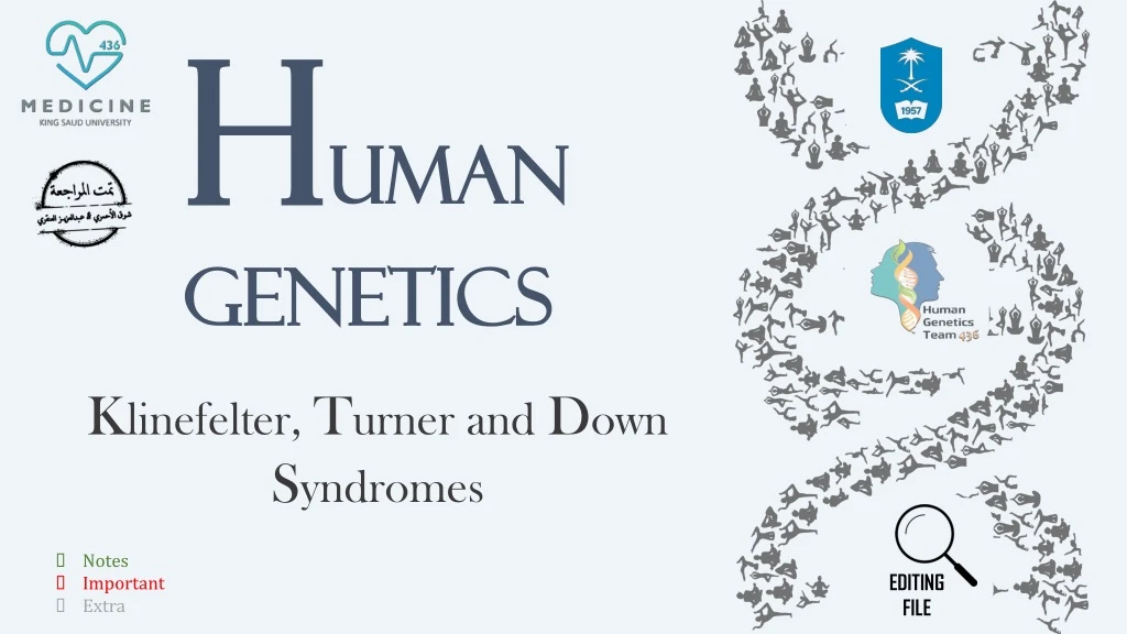
Human Genetics EDITING FILE Klinefelter, Turner and Down Syndromes • Notes • Important • Extra
Objectives Describe cell cycle and stages of Mitosis and Meiosis. Define nondisjunction and describe its consequences for meiosis and mitosis. Classify chromosomal abnormalities. Understand the common numerical chromosomal disorders: monosomy and trisomy. Understand the common numerical sex chromosome disorders: Down , Turner & Klinefelter syndromes.
RECALL(Extra) What is Chromosome? A thread-like structure of nucleic acids and protein found in the nucleus of most living cells, carrying genetic information in the form of genes. Human Chromosome: In humans cells, there is a set of 46 chromosomes organized in pairs -23 pairs per cell- and it is divided into two types: • 22 pairs of autosomes • One pair of sex chromosomes (Either XX or XY) Role of Chromosome: • Carry genetic material • Heredity • The intact set is passed to each daughter cell at every mitosis
Dr. Maram went through this quickly but she said (focus on the finale result of cell division). Cell Division The Cell Cycle: • Cellular components are replicated = Interphase • Cell distributes its contents into two daughter cells = Mitosis • G1 and G 2 = cell duplicates specific molecules and structures • S phase = cell replicates DNA • EXPLANATION : • The first step in the Cell Cycle is the INTERPHASE, which is divided into : G1 , S , G2 . • Interphase :the cell is growing and preparing to divide (it copies the DNA in preparation for mitosis), it spends most of its life in this phase. • G1: the chromosomes will UNTWISTED. • S: the genetic material will be replicated. • G2 : after replication they will be condensed again. • Following the interphase, the cell enter into MITOSIS which has FOUR phases : Prophase, Metaphase, Anaphase , Telophase. • The last step is the CYTOKINESIS : which is the division of the cytoplasm. The Cell Cycle (video)
Cell Division (Extra) Cell division is ( EXTRA but READ IT for better understanding) : The series of events that take place in a cell leading to its division and duplication of its DNA (DNA replication) to produce two daughter cells. • There are two distinct types of cell division which are: Meiosis Mitosis ① ❷ Mitosis VS Meiosis: • Mitosis is a vegetative division, whereby each daughter cell is genetically identical to the parent cell. • Meiosis is a reproductive cell division, whereby the number of chromosomes in the daughter cells is reduced by half to produce haploid gametes.
Dr Maram said events during the cells division are not important except METAPHASE Cell Division Mitosis in a human cell (Watch the Video for better understanding) Mitosis (Video) • It is IMPORTANT to know that NON DISJUNCTION (Not getting separated) happens at the ANAPHASE (the stage of separation), Either in Mitosis or Meiosis. • Chromosomes align during metaphase to be prepared for anaphase.
Cell Division Meiosis in human cell (Watch the Video for better understanding) Meiosis I Meiosis II • It is IMPORTANT to know that NON DISJUNCTION at the ANAPHASE 1 is more susceptible to have the abnormality. Meiosis (Video)
This table is IMORTANT (Focus on the Red) Comparison of Mitosis and Meiosis
Summary of the chromosome and chromatid number during Mitosis, Meiosis I & II in humans: “you Don’t have to worry about it just focus on the end product” (Haploid) Comparison of Mitosis and Meiosis
Non-disjunction Nondisjunction (Not coming apart): It is the failure of a chromosome pair to separate properly during meiosis I or of two chromatids of a chromosome to separate properly during meiosis II or mitosis. It can affect each pair each pair of chromosomes and is not a rare event. As a result, one daughter cell has two chromosomes or two chromatids and the other has none. (The aim of cell division is to get an equal number of chromosomes in each cell) • What do we mean by “not rare” event? • Those errors happen in daily basis during our life but since we have DNA repair, those errors cause no harm. Aneuploidy: It is the resulting cell of an imbalance of chromosomes due to nondisjunction. (A cell with the correct number of chromosomes is called a euploid cell). • Old age means slow cell division so nondisjunction increases. Examples: 47XXY (Klinefelter syndrome) Autosomal: Trisomy21 (Down Syndrome) Sex Chromosome: 45X (Turner syndrome)
Non-disjunction Non Disjunction in Meiosis 1 Non Disjunction in Meiosis 2 To Understand: Diploid (Diosomy) Haploid (monosomy) Nullisomy The Non Disjunction happened at which phase? Anaphase(in MEIOSIS which is the topic of this lecture mainly). Results(Please MEMORIZE them they are IMPORTANT): Non disjunction in meiosis 1 : 2 Diosomy (50%) , 2 nullisomy (50%) Non disjunction in meiosis 2 : 2 haploid , 1 nullisomy , 1 Diosomy
Non-disjunction Resulting Cells during normal segregation and Nondisjunction in Meiosis: • Normal : 4 Haploid gametes. • Nondisjunction in meiosis I : 2 gametes with diploid number of X chromosome and 2 gametes lacking X chromosome. • Nondisjunction in meiosis II: 2 gametes with haploid number of X chromosome, 1 gamete with diploid number of X chromosome, and 1 gamete lacking X chromosome. • When I ask you why does the defect occur ? • Because of excess or loss of expression of the genes . • How ? • When you have an extra chromosome it will lead to extra expression of the gene so you will have an extra feature and vice versa for the loss of chromosome)
Chromosome Anomalies Down Syndrome: • Also known as trisomy 21, is a genetic disorder caused by the presence of all or part of a third copy of chromosome 21. (Extra) • Karyotype: • Type of anomaly: • 47, XY, +21 (Trisomy 21) • Three copies of chromosome 21 Numerical anomaly in autosome (Extra) • Most cases arise from nondisjunction restricted to meiotic errors in the egg. • Mothers are the source of the extra chromosome in the majority of cases. • Advanced maternal age was significantly associated with both meiosis I (MI) and meiosis II (MII). • Nondisjunction occurred in MII, mothers were 15.1 times more likely to be ≥40 years compared to 8.5 times of nondisjunction in MI • The father contributing the extra chromosome in 15% of cases (i.e. Down syndrome can also be the result of nondisjunction of the father's chromosome 21). • A small proportion of cases are mosaic and these probably arise from a nondisjunction event in an early zygotic division =Mitotic. Down Syndrome (osmosis)
Chromosome Anomalies Down Syndrome Peak age for mothers with down syndrome babies • The incidence of trisomy 21 rises sharply with increasing maternal age. Meiosis II oocytes from younger and older women • As you can see the oocyte is disturbed with the increased age (basically the ovum also got aged), which increase the risk of the abnormality.
Chromosome Anomalies Down Syndrome Features: Dr Maram said read it with coffee Developmental delays (mental retardation) Life expectancy increased from 25 in 1983 to 60 today Head and facial malformations: (Small round face, protruding tongue = Sticks to the mouth floor) Low muscle tone = loose and floppy side Heart malformations Abnormalities of the extremities: (Short and broad hands, Stubby fingers), single deep crease across the center of the palm Impotency in males = Inability to sustain an erection sufficient for sexual intercourse or the inability to ejaculate
Sex chromosome imbalance is much less deleterious • (Please memorize the karyotype of each disorder) • 47,XYY Syndrome : • (May be without any symptoms). • Males are tall but normally proportioned. • 10 - 15 points reduction in IQ compared to sibs. • Klinefelter Syndrome (47,XXY) • Sex chromosome imbalance • Trisomy X (47,XXX) females: • It seems to do little harm. • Individuals are fertile and do not transmit the extra chromosome. • They do have a reduction in IQ comparable to that of Klinfelter males. • Turner Syndrome (45,X and variants)
Turner Syndrome (45,X and variants) : • (Now we moved to sex chromosome linked , Remember that Down Syndrome was Autosomal) Dr Maram said read it with coffee • Monosomy of sex chromosome: (Monosomy X: 45, XO) i.e. only one X chromosome is present. • Note that it can be written 45,X or 45,XO ( the O is just to show that there is a missing chromosome). • Occurrence – 1 in 2500 live female births. • The only viable monosomy in humans. • Individuals are genetically female, not mature sexually and sterile. Turner syndrome Turner Syndrome (Video)
Features of Turner Syndrome: Dr Maram said read it with coffee Lack of ovarian development (Streak ovaries) = No ovaries (infertile) Normal life span Neck abnormalities (webbed neck) Turner syndrome Increased risk of osteoporosis, cardiovascular anomalies e.g. constriction of aorta and hypertension Short stature, Broad chest, Low hairline Skeletal disorders (e.g. scoliosis, dislocated hips/elbows) No developmental delays, Normal intelligence
Features of Turner Syndrome Continued… • Cardiovascular : • Bicuspid aortic valve. • Coarctation of aorta. • Thoracic aortic aneurysm (aortic root dilatation). • Skeletal : • Short stature . • Short 4th metacarpal/metatarsal bone (± short 3rd and 5th). • Osteoporosis (due to lack of estrogen). • Scoliosis. Turner syndrome • Reproductive: • Women with Turner syndrome are almost universally infertile.
Klinefelter Syndrome • 1 in 1,100 births. • 47 chromosomes. • Karyotype: 47, XXY. • Very rarely more extreme forms of Klinefelter syndrome occur where the patient has 48, XXXY or even 49, XXXXY karyotype. (These individuals are generally severely retarded). Klinefelter Syndrome Klinefelter Syndrome (Osmosis)
Normal life span DrMaram said read it with coffee Features of Klinefelter Syndrome: Deficits in attention, auditory processing and social skills. Sexually underdeveloped & infertile* (no spermatogenesis) * In some cases testicular function is preserved Klinefelter Syndrome Delays in speech and motor skills Tall Low mental ability (slight reduction in IQ, but usually normal intelligence) Sparse facial and body hair
Features of Klinefelter Syndrome Continued… Dr Maram said read it with coffee Longer fingers and arms Delicate skin Gynecomastia and other feminine body characteristic Increased risk of autoimmune disorders, breast cancer, osteoporosis, leg ulcers, depression, and dental problems Klinefelter Syndrome Treatment: • Includes testosterone therapy and assisted learning.
When to do a chromosomal test: Tests • IMPORTANT and you have to know : the most important Tests are? FISH and Karyotyping. • During pregnancy, ultrasound can be used to detect down syndrome by observing special signs in the fetus such as increase the thickness of back of the baby neck.
Rapid Aneuploidy Screening by Fluorescence in situ hybridization (FISH): • Available on amniocentesis sample. • Uncultured amniocytes. • FISH probes for X,Y, 21 • Result in 24-48 hours. • Proceed onto full karyotype (11-14 days). Tests New techniques: • Cell-free fetal DNA from maternal plasma – at 6-8 weeks of gestation: • It is a non-invasive prenatal diagnostic tool for chromosomal aneuploidy. It can be used to determine the fetus sex–: look for presence of Y chromosome material. • Quantitative Fluorescence PCR (qf PCR): • isable to measure number of copies of a chromosome – used for trisomy screening.
Normal human karyotype is 46,XY or 46,XX • Chromosome abnormalities can be numerical or structural. • Numerical abnormalities include aneuploidy and polyploidy. • In monosomy or trisomy, a single extra chromosome is absent or present, usually as a result of nondisjunction in the 1st or 2nd meiotic division. • Structural abnormalities include translocations, inversions, deletions, isochromosome & rings. Take Home Messages (don’t Skip!!)
Cell cycle include interphase ( where Cellular components are replicated ) mitosis (type of cell division occur in somatic cells ). • Mitosis > One Division>Two daughter cells per cycle(diploid )>somatic cells. • Meiosis >Two Divisions>Four daughter cells per cycle (Haploid)> germ line cells . SUMMARY
Q1. Non-Disjunction Defect in the meiotic cell division happens at which phase? Prophase Metaphase Anaphase Telophase Q4.Which one of the following tests is the best for detecting a chromosomal abnormality? FISH Karyotyping PCR A & B Q2. Meiosis Occurs in which one of the following cells ? A. Somatic cells B. Germline cells C. Ovum and sperm D. B & C • Q5.Which one of the following chromosomal disorders correlates with the karyotype 47,XXY ? • Klinefelter Syndrome • Down Syndrome • Turner Syndrome • Trisomy X Test Yourself !! Q6. which one of the following is the result of non-disjunction in meiosis 1 ? A. 2 haploid , 1 nullisomy , 1 Diosomy B. 2 Diosomy , 2 nullisomy C. 4 Haploid D. 3 Haploid , 1 nullisomy Q3. A 16 years old Girl presented to the hospital complaining of delayed puberty, on examination the doctor noticed the that patient is short with a webbed neck, Chromosomal karyotyping showed 45,X . What is the diagnosis? Klinefelter syndrome Down Syndrome Turner Syndrome Constitutional delayed puberty Answer key: 1:C 2:D 3:C 4:D 5:A 6:B
“One page can change you, many will change the world” Team Leaders: Mohammed Habib. Aseel Badukhon. Special thanks to: Reema Al-Barrak Reference Male’s and Female’s Slides Humangenetics436 @436Genetics Your feedback?