Ankle Fracture Update
Ankle Fracture Update. OTA Resident Core Curriculum Lecture Series Updated November 2010. Matt Graves, M.D. University of Mississippi Medical Center. Objectives. Following this session, you should be able to: State the indication to fix isolated fibular fractures.

Ankle Fracture Update
E N D
Presentation Transcript
Ankle Fracture Update OTA Resident Core Curriculum Lecture Series Updated November 2010 Matt Graves, M.D. University of Mississippi Medical Center
Objectives • Following this session, you should be able to: • State the indication to fix isolated fibular fractures. • Define the specific articular pathology associated with SA and PAB fractures. • List the 3 common posterior malleolar fracture patterns. • State the indication to fix posterior malleolar fractures. • Enumerate the ways to ensure syndesmotic reduction.
Recommendations to Improve Retention of this Material • Write down the objectives • Search for the answers to the objectives in the powerpoint talk [hint- look for blue boxes] • Test yourself at the end by reviewing the objectives • Watch the show on “normal view” and look at the notes at the bottom of the slides. They will provide guidance to the progression of logic and sources of information. Classic references are listed throughout. Annotated recent references are listed at the end.
Outline • Evaluation: Clinical & Radiographic • Classification: Lauge-Hansen • Specific Problem Areas: Posterior Malleolus and Syndesmosis • Surgical Goals • Outcome
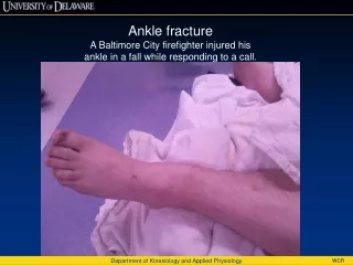
Evaluation:Clinical HISTORY PHYSICAL EXAM Skin Nerves Vasculature Pain Deformity • Mechanism • Timing • Soft-tissue injury • Bone quality • Comorbidities • Associated Injuries
Evaluation: RadiographicAnteroposterior View • Tibiofibular overlap ~ 10mm • Tibiofibular clear space<5mm • Talar tilt Comparison Radiograph?
Evaluation: RadiographicMortise View 10 degrees internal rotation of 5th MT with respect to a vertical line Goergen JBJS 1977
Evaluation: RadiographicMortise View • Medial joint space • Talocrural angle: <8 or >15 degrees • Tibia/fibula overlap:>1mm Comparison Radiograph?
Evaluation: RadiographicMortise View FIBULAR LENGTH: 1. Shenton’s Line of the ankle 2. The dime test Weber SICOT 1981
Evaluation: RadiographicLateral View • PM • Talar subluxation • Distal fibular translation &/or angulation • Syndesmotic relationship • Associated or occult injuries • Lateral process talus • Posterior process talus • Anterior process calcaneus
Evaluation: RadiographicOther Imaging Modalities • Stress Views • Gravity • Manual • CT • Articular involvement • Posterior malleolus • MRI • Ligament and tendon injury • Talar dome lesions • Syndesmosis injuries
Outline • Evaluation: Clinical & Radiographic • Classification: Lauge-Hansen • Specific Problem Areas: Posterior Malleolus and Syndesmosis • Surgical Goals • Outcome
Lauge-Hansen • Cadaveric study • First word: position of foot at time of injury • Second word: force applied to foot relative to tibia at time of injury Types: SER SA PER PA
Lauge-Hansen • Several stages per type • Imperfect system: • Not every fracture fits exactly into one category • Even mechanismspecific pattern has been questioned • Inter and intraobserver variation not ideal • Still useful and widely used
Supination-External Rotation Stage 1- AITFL Stage 2- Fibula fx Stage 3- PITFL or PM fx Stage 4-Deltoid or MM fx 70% of ankle fractures
Supination-External Rotation Stage 2: Stable Stage 2 Lateral Injury: classic posterosuperioranteroinferior fibula fracture Medial Injury: Stability maintained Standard: Closed management Kristensen Acta Orthop Scand 1985
Supination-External Rotation Stage 4: Unstable Stage 4 Lateral Injury: classic posterosuperioranteroinferior fibula fracture Medial Injury: medial malleolar fracture &*/or deltoid ligament injury Standard: Surgical management *Tornetta JBJS 2000
SER-2 vs SER-4: How to Decide? • Michelson. Clin Orthop Rel Res 2001 • DeAngelis Poster OTA 2003 • Tornetta. Poster AAOS 2004 • McConnell JBJS 2004 • Egol JBJS 2004 • Schock Presentation OTA 2006 • Zeni Presentation OTA 2006 • Park J Orthop Trauma 2006 GOAL: TO EVALUATE DEEP DELTOID [i.e. INSTABILITY] MEDIAL TENDERNESS METHOD: MEDIAL SWELLING MEDIAL ECCHYMOSIS STRESS VIEWS- GRAVITY OR MANUAL
Gravity Stress Exam Michelson et al. CORR 387: 178-82, 2001.
versus • Both are effective • Gravity stress requires XR education. • Manual stress requires time and more radiation exposure. Schock et al. JBJS 89B: 1055-59, 2007.
SER-2 vs. SER-4: How To Decide? Indication to fix isolated fibular fractures
Decision-Tree:Understand the Logic • Assumptions: • Fibular fractures associated with a stable ankle mortise heal without significant functional consequence. • Fibular fractures associated with an unstable ankle mortise heal with significant functional problems…because instability allows for talar shift.
Decision Tree:Understand the Logic Stress View Splintage
Decision-Tree:Understand the Logic • Does a Positive Ankle Stress Test Indicate the Need for Operative Treatment? • MRI to evaluate all patients with lateral malleolar fracture and positive stress test (n=21). • If deep deltoid partially intact nonop treatment • Good clinical outcomes. • OTA Annual Meeting. Foot & Ankle Section. Paper #24, 2006.
Indication to fix isolated fibular fractures Choose a technique to evaluate stability. Base your decision to operate on your findings and the risk:benefit ratio.
Supination Adduction • Stage 1: transverse Weber A or B fibula • Stage 2: vertical medial malleolus
Supination Adduction: Stage 2 Lateral Injury: transverse fibular fracture at/below level of mortise Medial injury: vertical shear type medial malleolar fracture BEWARE OF IMPACTION McConnell J Orthop Trauma 2001
Supination Adduction: Stage 2 • Important to restore: • Ankle stability • Articular congruency- including medial impaction
SAD • Consider anteromedial approach • Marginal impaction reduction +/- grafting • Medial antiglide plate Specific articular pathology associated with SA
Pronation-External Rotation • Stage 1 - deltoid or medial malleolus • Stage 2- AITFL and IO membrane • Stage 3 – spiral Weber C fibula • Stage 4 – PITFL or posterior malleolus
Pronation External Rotation: Stage 4 Medial injury: deltoid ligament tear &/or transverse medial malleolar fracture Lateral Injury: spiral proximal lateral malleolar fracture HIGHLY UNSTABLE…SYNDESMOTIC INJURY COMMON
PER • Tibia radiograph • Syndesmostic disruption expected • Restore: • Fibular length and rotation • Ankle mortise • Syndesmotic stability
Pronation-Abduction • Stage 1 – transverse MM • Stage 2 – PITFL or PM fracture • Stage 3 – compression bending fibula fracture
Pronation-Abduction Medial injury: tranverse to short oblique medial malleolar fracture Lateral Injury: comminuted impaction type lateral malleolar fracture
PAB • Medial malleolar fixation drives stability. Go there 1st. • Fibular comminution length stable construct? • Stress the syndesmosis last JBJS 89A: 276-81, 2007
PAB Specific articular pathology associated with PAB
Outline • Evaluation: Clinical & Radiographic • Classification: Lauge-Hansen • Specific Problem Areas: Posterior Malleolus and Syndesmosis • Surgical Indications and Goals • Outcome
Posterior Malleolus Fractures Function: Stability- prevents posterior translation of talus & enhances syndesmotic stability Weight bearing- increases surface area of ankle joint
Posterior Malleolus Fractures: Radiographic Evaluation • Fracture pattern: • Variable • Difficult to assess on standard lateral radiograph • External rotation lateral view [Decoster FAI 2000] • CT scan [Haraguchi JBJS 2006]
Posterior Malleolus Fracture: Radiographic Evaluation • Indication for fixation: > 25% joint surface on lateral • Problem: Fragment size hard to determine on lateral view • Reason: Fracture orientation not purely in coronal plane • Nearly always associated with the pull of the posterior tib-fib ligament • larger laterally than medially • obliquely oriented • involves the incisura Haraguchi et al. JBJS 2006 …but other fracture patterns have also been defined
Posterior Malleolus Fracture 67% 19% Type I- posterolateral oblique type Type II- medial extension type 14% Type III- small shell type 3 common PM fracture patterns Haraguchi et al. JBJS 2006
Posterior Malleolus Fractures: Indications for Fixation • Stability • Posterior translation of talus* • ER of talus [syndesmotic widening] • Articular congruence • Stress = Force/Area • Excessive stressposttraumatic arthritis • Maximize area for stress distribution** *fibula and anterior tibiofibular ligament act as primary restraint [Raasch JBJS 1992] **contact stress changes significantly with posterior malleolar size >33% [Hartford CORR 1995]
Posterior Malleolus Fracture: Fixation • Screws • Plates
Syndesmotic Injury FUNCTION: Stability- resists external rotation, axial, & lateral displacement of talus Weight bearing- allows for standard loading
Syndesmosis IF INSTABILITY PRESENT OPERATIVE INTERVENTION OBTAINING & MAINTAINING ANATOMIC REDUCTION REDUCES LONG TERM DISABILITY & IMPROVES sMFA Leeds JBJS 1984 Weening JOT 2005
Syndesmosis:Instability • How do you determine if instability is present? • Manual Stress Test • When do you perform the manual stress test? • After you have fixed the other indicated components of the fracture