Download
1 / 39
390 likes | 407 Vues
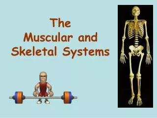
This chapter discusses the skeletal and muscular systems in vertebrates, including bone structure, bone density, joint types, and muscle structure. It also covers the role of the nervous system in muscle contraction.

E N D
Endoskeleton • All vertebrates • Fins or limbs attach to skeleton at pectoral and pelvic girdles Generalized mammal Figure 37.6dPage 642 pelvic girdle pectoral girdle
SKULL PECTORAL GIRDLES AND UPPER EXTREMITIES cranial bones facial bones clavicle RIB CAGE scapula sternum humerus ribs radius ulna VERTEBRAL COLUMN vertebrae phalanges carpals intervertebral disks metacarpals PELVIC GIRDLE AND LOWER EXTREMITIES pelvic girdle femur patella tibia fibula tarsals phalanges metatarsals
Long Bone Structure • Compact bone • Spongy bone • Central cavity contains yellow marrow nutrient canal contains yellow marrow compact bone tissue spongy bone tissue Fig. 37.12 (1)Page 652
Compact Bone Structure • Mature compact bone consists of many cylindrical Haversian systems Haversian system blood vessel outer layer of dense connective tissue spongy bonetissue Fig. 37.12 (2)Page 652 compact bone tissue
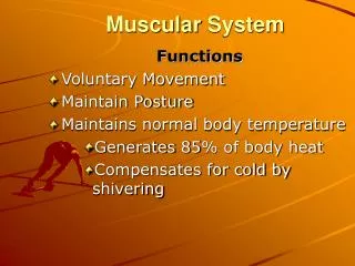
Functions of Bone • Interact with muscle to enable movement • Support and anchor muscles • Enclose and protect internal organs • Store calcium and phosphorus • Produce blood cells
Bone Marrow • Yellow marrow • Fills the cavities of adult long bones • Is largely fat • Red marrow • Occurs in spongy bone of some bones • Produces blood cells
Bone Remodeling • In adults, bone building and bone breakdown continue constantly • Osteoblasts deposit bone • Osteoclasts secrete enzymes that degrade it • Remodeling adjusts bone strength and helps maintain blood calcium levels
Embryo: cartilage model of future bone in embryo Fetus: blood vessel invades model; osteoblasts start producing bone tissue; marrow cavity forms Newborn: remodeling and growth continue; secondary bone-forming centers appear at knobby ends of bone Adult: mature bone Fig. 37-9, p.645
Cartilage model of future bone in embryo Blood vessel invades model; osteoblasts start producing bone tissue; marrow cavity forms Remodeling and growth continue in newborn; secondary bone-forming centers appear at knobby ends of bone Mature adult bone Stepped Art Fig. 37-9, p.645
Bone Density • Exercise can increase bone density • Osteoporosis is a decrease in bone density • May occur when the action of osteoclasts outpaces that of osteoblasts • May also occur as a result of inability to absorb calcium
Bone Density Fig. 37-10a, p.645
Bone Density Fig. 37-10b, p.645
Joints • Areas of contact or near contact between bones • Fibrous joints • Short connecting fibers join bones • Synovial joints • Move freely; ligaments connect bones • Cartilaginous joints • Straps of cartilage allow slight movement
Tendons Attach Muscle to Bone muscle tendon bursae synovial cavity Figure 37.13bPage 647
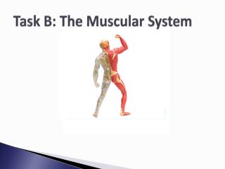
Skeletal Muscle • Bundles of striped muscle cells • Attaches to bone • Often works in opposition biceps triceps Figure 37.11Page 646
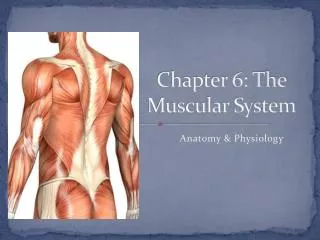
triceps brachii triceps brachii biceps brachii deltoid pectoralis major pectoralis major trapezius serratus anterior serratus anterior external oblique external oblique latissimus dorsi rectus abdominus rectus abdominus gluteus maximus adductor longus adductor longus biceps femoris biceps femoris sartorius sartorius quadriceps femoris quadriceps femoris gastrocnemius gastrocnemius tibialis anterior tibialis anterior Figure 37.13Page 647
Skeletal Muscle Structure • A muscle is made up of muscle cells • A muscle fiber is a single muscle cell • Each fiber contains many myofibrils myofibril Figure 37.14aPage 648
Sarcomere A myofibril is made up of thick and thin filaments arranged in sarcomeres sarcomere sarcomere sarcomere sarcomere Z line Z line Z line Figure 37.14cPage 648
Muscle Microfilaments Thick filaments • Composed of myosin • Each myosin molecule has tail and a double head Thin filaments • Like two strands of pearls twisted together • Pearls are actin • Other proteins in grooves in filament Figure 37.14d-ePage 648
Sliding-Filament Model • Myosin heads attach to actin filaments • Myosin heads tilt toward sarcomere center, pulling actin with them Fig. 37.20c-gPage 657
Nervous System Controls Contraction • Signals from nervous system travel along spinal cord, down a motor neuron • Endings of motor neuron synapse on a muscle cell at a neuromuscular junction
Role of Calcium in Contraction • T tubules in the sarcoplasmic reticulum relay signal • Calcium ions are released
Troponin and Tropomyosin • Lie in groove in actin filament • When muscle is relaxed, tropomyosin blocks myosin binding site troponin myosin binding site blocked actin Figure 37.17bPage 651
Troponin and Tropomyosin • When troponin binds calcium ions, it changes shape and moves tropomyosin • Cross-bridge formation and contraction can now proceed myosinhead actin Figure 37.17ePage 651
a Actin molecule and associated proteins myosin binding site blocked b Transverse section of (a). Red dots are calcium ions bound to troponin (green) Fig. 37-17a,b, p.651
c Influx of calcium ions; troponin binds additional calcium d Troponin changes shape, moves away from myosin binding site. Fig. 37-17c,d, p.651
myosinhead e Binding site is now exposed; actin is free to bind myosin head myosinhead f Cross-bridge forms between actin, myosin Fig. 37-17e,f, p.651
Contraction Requires Energy • Muscle cells require huge amounts of ATP energy to power contraction • The cells have only a very small store of ATP • Three pathways supply ATP to power muscle contraction
Muscle Tension • Mechanical force a contracting muscle exerts on an object • For a muscle to shorten, muscle tension must exceed the load that opposes it • The load may be the weight of an object or gravity’s pull on the muscle
Two Types of Contraction contracted muscle can’t shorten contracted muscle can shorten isotonic contraction isometric contraction Figure 37.20a-bPage 652
Motor Unit • One neuron and all the muscle cells that form junctions with its endings • When a motor neuron is stimulated, all the muscle cells it supplies are activated to contract simultaneously • Each muscle consists of many motor units
Muscle Fatigue • An inability to maintain muscle tension • Occurs after a period of tetanic contraction • Different types of muscle show different fatigue patterns
triceps brachii triceps brachii biceps brachii deltoid pectoralis major pectoralis major trapezius serratus anterior serratus anterior external oblique external oblique latissimus dorsi rectus abdominus rectus abdominus gluteus maximus adductor longus adductor longus biceps femoris biceps femoris sartorius sartorius quadriceps femoris quadriceps femoris gastrocnemius gastrocnemius tibialis anterior tibialis anterior Figure 37.13Page 647