Download
1 / 67
830 likes | 2.08k Vues
Head & Neck Masses including Salivary Glands. Dr Imtiaz M Qazi. Highlights of Neck A natomy. Prominent landmarks Triangles of the neck Lymphatic levels. Prominent L andmarks. Hyoid bone Thyroid cartilage Cricoid cartilage Trachea Sternomastoid M . Triangles of the neck.

E N D
Head & Neck Massesincluding Salivary Glands Dr Imtiaz M Qazi
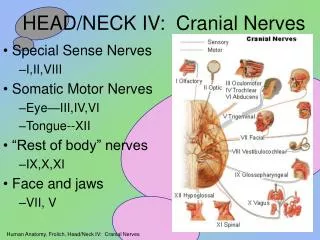
Highlights of Neck Anatomy • Prominent landmarks • Triangles of the neck • Lymphatic levels
Prominent Landmarks • Hyoid bone • Thyroid cartilage • Cricoid cartilage • Trachea • SternomastoidM
Triangles of the neck • Sternomastoid ÷ neck into: • Anterior triangle • Posterior triangle
Anterior triangle ÷ by digastric & sup. belly of omohyoid into: • submandibular • submental • carotid • muscular • Posterior triangle ÷ by inf. belly of omohyoid into: • occipital • supraclavicular
Adults v/s Children • Children 90% of neck masses are benign • Asymptomatic neck masses - adults >40 yrs old consider malignant until proven otherwise • 80% of non-thyroid and non-salivary gland masses are neoplastic (80% of which are malignant)
Branchial Cleft Cyst • Pathophysiology: Developmental alterations of the branchial apparatus • Cysts, sinuses, or fistulas • Cysts: 10-40 yrs of age • Sinuses and Fistulas : 1st decade
First Branchial Cleft Cyst • External opening • Type 1 • Usually Preauricular cyst • Duplicated EAC • Ectodermal elements only • Complete excision after infection has resolved
First Branchial Cleft Cysts • Type II • More common • Submandibular Cyst/ tract below angle of mandible and through the parotid in variable position to CN VII • Tract runs from the neck to the EAC or middle ear • Surgical excision- superficial parotidectomy
Second Branchial Cleft Cysts • Most Common (90%) branchial anomaly – failure of obliteration of cervical sinus of His • Painless, fluctuant mass in anterior triangle along ant. border of SCM • Can occur at carotid bifurcation or parapharyngeal space • Upper & middle 3rdjunction of SCM, deep to platysma, lateral to IX, X, XII, between the internal and external carotid and terminate in the tonsillar fossa • Surgical treatment may include tonsillectomy
Third Branchial Cleft Cysts • Rare (2%) • Masses low in the anterior neck, more often on the left side • Internal opening at the pyriform sinus • Often track through the upper pole of the thyroid
Fourth Branchial Cleft Cysts • Extremely rare • Cyst anterior to inferior portion of SCM • May have opening located near the apex of the pyriform sinus, fistula or sinus tract that travels between the superior and inferior laryngeal nerve
Thyroglossal Duct Cyst • Most common congenital midline mass • Failure of complete obliteration of the thyroglossal duct • Elevates with tongue protrusion • Commonly at the level of the hyoid • Ectopic thyroid tissue vs. thyroglossal duct cyst • Ultrasound • Radioisotope scan • Sistrunk’s Operation
Cervical Thymic Cysts Commonly in the lower neck, but anywhere from the pyriform sinus to the chest Failure of involution of the cervical thymo pharyngeal ducts (remnant of 3rdparyngeal pouch) Firm, mobile mass found in the lower aspect of the neck CXR, CT scan, Biopsy Treatment: Excision
Dermoid Cysts • Tissue from all three germinal layers • Midline, paramedian, painless • Misdiagnosed as Thyroglossal Duct Cysts • Do not elevate with tongue protrusion • Complete surgical excision to prevent recurrence
Teratoma • All three germ cell layers, but foreign to the site of presentation • Mature vs. immature • Larger midline masses • Rarely present after the first year of life • 20% associated maternal polyhydramnios • Unlike adult teratomas, they rarely demonstrate malignant degeneration • Surgical excision
Laryngocele • Enlarged laryngeal saccule • distension and entrapment of air • Classified as internal, external, or combined
Internal Laryngocele • Confined to larynx, usually involves the false cord and aryepiglottic fold • Hoarseness and respiratory distress vs. neck mass
External and Combined Laryngoceles • Soft, compressible, lateral neck mass that distends with increases in intralaryngeal pressures • Through the thyrohyoid membrane at the entrance of the Superior Laryngeal Nerve • CT scan
Lymphangioma Soft, painless, multiloculated, compressible neck mass 80% in post triangle 65% at birth, 90% by 3 yrs 40% present with airway compromise Transilluminates US, CT, MRI Spontaneous regression rare: 8 - 15% Sclerotherapy v/s Surgical excision Recurrence : 10-50%
Cystic Hygroma • Described by Wernher (1843) • Head and neck (75%) • L>R • 1/6000-16000 • African american • M=F
Haemangioma • Hemangioma • Most common tumor of infancy • Less than 33% present at birth • Rapid growth then a period of quiescence followed by involution • If associated with stridor R/O Subglottichaemangioma • Treatment
Vascular malformations • Nevus flammeus vs. port wine stain • Sturge-Weber Syndrome • Venous Malformations • Lips and cheeks • Expand with jugular venous congestion • Intraosseous “soap bubble” appearance • AVM • High flow • Thrill/bruit • Pain, ulceration, bleeding and pulsatile tinnitus
Plunging Ranula • Uncommon • Simple - unilateral cystic lesion • Visible under the tongue • Plunging - through mylohyoid • Cyst aspirate- high protein, amylase levels • CT scan/MRI • Treatment is intra-oral excision to include the sublingual gland of origin
Fibromatosis colli • Torticollis with firm mass on the SCM • Noted at birth or within 1st few weeks • Inflammatory lesion of unknown etiology with muscle replacement by fibrosis • Range of motion exercises • Myoplasty of the SCM only if refractory to PT
Carotid Body Tumour Chemodactoma/Paraganglioma Paraganglionic cells of carotid body at bifurcation of the carotid artery Highly vascular 4th – 5th decades of life Female preponderance 10% bilateral Radiological feature: Lyre Sign Shamblin Classification: Group 1 – minimally attached to vessels & easily removed Group 2 – partially surround vessels, more adherent to the adventitia Group 3 – adherent to the entire surface of carotid bifurcation, resection impossible
Pharyngeal /Zenker’sDiverticulum Out-pouching of the pharynx between the muscles at the junction of the pharynx and oesophagus Elderly pts > 70yrs Dysphagia, regurgitation, chronic cough, aspiration, and weight loss Diagnosis: Ba Swallow Treatment: Surgical via an endoscopic or external cervical approach and should include a cricopharyngealmyotomy
Neoplasms BENIGN • 1. Vascular • Hemangioma • Lymphangioma • arteriovenous malformation • Aneurysm • 2. Chemodectoma • 3. Neural—neurofibroma, schwannoma • Parapharyngeal space with tonsillar displacement • 4. Lipoma • >35 y/o • On CT : Fat:air density • 5. Fibroma • 6. Miscellaneous—fibromatosis
Neoplasms MALIGNANT • Neck primary • Lymphoma • All age groups • Sarcoma • Rhabdomyosarcoma: pedia • Osteosarcoma: Adults • Thyroid carcinoma • Pediatric: M>F ; malignancy • Adult: F>M; benign conditions • Salivary gland carcinoma • Pain, rapid growth, CN VII symptoms, skin fixation
Neoplasms • Metastatic • Head and neck • Infraclavicular primary • Leukemia
Inflammatory/Infectious • CERVICAL LYMPHADENITIS • Most common cause of neck masses • Usually don’t require intervention (reactive) • VIRAL (URTI) • BACTERIAL • Granulomatous disease • Typical – adults • Atypical - pediatrics • Lymph node hyperplasia due to AIDS • Biopsy :single enlarged (>3 cm) node
Conclusion Neck masses are very common Approach with History and Physical exam will commonly lead to the correct diagnosis Laboratory and radiographic exams are confirmatory To know where you are heading better look into the origin (embryology) A nodal mass is almost always not the primary mass Nodal spread for infections and neoplasm (better familiarize them)
LOCATION • Important!!! • Congenital and developmental mass • Consistent location • Malignancy and inflammation – same spread • (lymphatics)
Some pearls: • 90% of adult neck masses are malignant • 90% of pediatric neck masses are infectious in nature • Know your anatomy then develop a differential diagnosis • Close observation • Generally, one course of a broad spectrum antibiotic is acceptable then …..
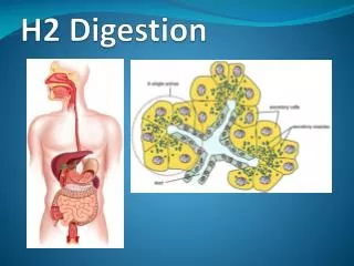
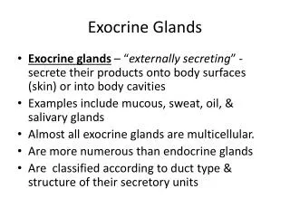
Salivary Glands • Major salivary gland a. Parotid gland b. Submandibular gland c. Sublingual gland 2. Minor salivary gland 600 – 1,000 minor salivary gland distributed throughout the mucosa of the upper aerodigestive tract (more common in the soft and hard palate)
Salivary Gland Tumors 80% of salivary gland tumor occur in the parotid 10 – 15% in the minor salivary gland 5 – 10% in the submandibular gland 80% of the parotid tumor are benign The most common is pleomorphicadenoma 50% of the submandibular gland tumor are benign 30% of the minor salivary gland are benign
Salivary Gland Tumours From: Hanna EY, Lee S, Fan CY, Suen JY. Chapter 60: Salivary Gland Physiology. In: Cummings et al. (Eds.). Cummings: Otolaryngology – Head and Neck Surgery, 4th Ed. 1998, Mosby, Philadelphia, PA.
Benign Salivary Gland Tumours 1. Pleomorphicadenoma (benign mixed tumor) 2. Warthin’stumor (papillary cyst adenoma lypmhomatosum) Monomorphicadenoma a. Basal cell adenoma b. Canalicular adenomas c. Oncocytoma d. Myoepitheliomas 3. Granular cell tumor 4. Hemangioma
Malignant Salivary Gland Tumours 1. Mucoepidermoid carcinoma – 40% 2. Adenoid cystic carcinoma – 10% 3. Acinic cell carcinoma – 10 – 15 % of It is considered a low-grade tumor 4. Malignant mixed tumor - 7% It is considered a high-grade malignancy 5. Polymorphous low grade adenocarcinoma– 10% It is a low-grade variant of adenocarcinoma 6. Adeno carcinoma – 10% It is a high-grade with poor prognosis 7. Squamous cell carcinoma – 4% It is high-grade, more common in elderly patients, and can confused with high-grade mucoepidermoidcarcinoma
Pleomorphic Adenoma • Most common of all salivary gland neoplasms • 70% of parotid tumors • 50% of submandibular tumors • 45% of minor salivary gland tumors • 6% of sublingual tumors • 4th-6th decades • F:M = 3-4:1
Pleomorphic Adenoma • Slow-growing, painless mass • Parotid: 90% in superficial lobe, most in tail of gland • Minor salivary gland: lateral palate, submucosal mass • Solitary vs. synchronous/metachronous neoplasms
Pleomorphic Adenoma • Treatment: complete surgical excision • Parotidectomy with facial nerve preservation • Submandibular gland excision • Wide local excision of minor salivary gland • Avoid enucleation and tumor spill
Warthin’s Tumor • AKA: papillary cystadenomalymphomatosum • 6-10% of parotid neoplasms • Older, Caucasian, males • 10% bilateral or multicentric • 3% with associated neoplasms • Presentation: slow-growing, painless mass
Oncocytoma • Rare: 2-3% of benign salivary tumors • 6th decade • M:F = 1:1 • Parotid: 78% • Submandibular gland: 9% • Minor salivary glands: palate, buccal mucosa, tongue • Enlarging, painless mass
Mucoepidermoid Carcinoma • Most common salivary gland malignancy • 5-9% of salivary neoplasms • Parotid 45-70% of cases • Palate 18% • 3rd-8th decades, peak in 5th decade • F>M • Caucasian > African American
Mucoepidermoid Carcinoma • Presentation • Low-grade: slow growing, painless mass • High-grade: rapidly enlarging, +/- pain • **Minor salivary glands: may be mistaken for benign or inflammatory process • Hemangioma • Papilloma • Tori