Download
1 / 30
300 likes | 492 Vues
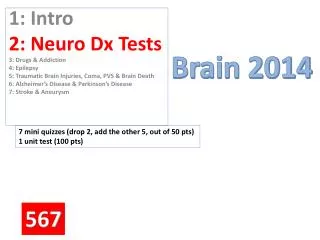
Brain 2014. 7 mini quizzes (drop 2, add the other 5, out of 50 pts) 1 unit test (100 pts). 1: Intro 2: Neuro Dx Tests 3: Drugs & Addiction 4: Epilepsy 5: Traumatic Brain Injuries, Coma, PVS & Brain Death 6 : Alzheimer’s Disease & Parkinson’s Disease 7 : Stroke & Aneurysm. 567.

E N D
Brain 2014 7 mini quizzes (drop 2, add the other 5, out of 50 pts) 1 unit test (100 pts) 1: Intro 2: NeuroDx Tests 3: Drugs & Addiction 4: Epilepsy 5: Traumatic Brain Injuries, Coma, PVS & Brain Death 6: Alzheimer’s Disease & Parkinson’s Disease 7: Stroke & Aneurysm 567
On notebook paper • In 3-ring binder, BRAIN section • USE ONE FULL SIDE OF PAPER • Create this table
Neurological Diagnostic Tests MQ#2 DAY 3/6
First, let’s understand… • Soft tissue vs hard tissue (brain, cranium, carotid artery, cerebrum, cerebellum, brain stem) • “normal” xrays???
1. Computerized Tomography (CT) Scan • Aka CAT (A=axial) • Combines Xrays & computers • Visualizes soft & hard tissue • Can be compared to slicing a loaf of bread • Can involve contrast (dye) sometimes (either PO or IV) • Can see internal organs, tumors, blood vessels, joints, etc. http://www.youtube.com/watch?v=uHu9aa0QDiE
Why Done: • Pinpoint a tumor, fracture, infection or blood clot, injuries, bleeding • Guide procedures (ex. surgery, biopsy) • Brain PLUS
Risks • Radiation • Allergic to contrast Tumor Probable S/S???
What you can expect • Remove all metal • Fasting, @ times • Painless • Only a few minutes • “Donut hole”; claustrophobic for some 57
2. Magnetic Resonance Imaging (MRI) M = use of magnets (NOT xrays) R = vibrations, bouncing, echoing I = pictures How it works: Magnets react with body’s hydrogen to make pic’s 6 http://www.youtube.com/watch?v=DZTXa4qerI4 3 4?
Why done The test of choice for visualizing soft tissue (brain PLUS): • Organs • Ligaments • Circulatory system • Brain & spinal cord
Risks • Newly-implanted metal body parts • Otherwise, fairly risk-free
What to Expect • Remove ALL metal & credit cards • Very tight donut hole (claustrophobic, anyone?) • Painless • Must lie VERY still • Loud banging / headphones
There have been reports of people with tattoos or permanent makeup or who experience swelling or burning in the tattooed areas during magnetic resonance imaging (MRI). This seems to occur only rarely and apparently without lasting effects. There have also been reports of tattoo pigments interfering with the quality of an MRI image. This seems to occur mainly when a person with permanent eyeliner gets an MRI of the eyes. Mascara may produce a similar effect, but it can easily be removed. Why these problems happen is unclear. It's possible they result from an interaction with the metallic components of some pigments. The risks of avoiding an MRI when your doctor has recommended one are likely to be much greater than the risks of complications from a tattoo. Instead of avoiding an MRI, people who have tattoos or permanent makeup should tell the radiologist or technician in order to take precautions to avoid complications
A. CT B. MRI • As a pair or trio… • Answer these 6 ?s • Provide rationale for your answers
A. CT B. MRI • Which is OK for pregnant lady? Why? • Which exposes the pt to fairly high dose of radiation? Why? • Which does NOT use radiation? Why? • Which is safer for (growing) children? Why? • Which is better for an obese person? Why? • Which is better for someone who’s extremely claustrophobic? Why?
3. EEG • Electro EncephaloGram • Records brain’s electrical activity
Why Done • To dx seizure disorders • Also, sometimes sleep disorders Epilepsy!!!
Risks • Can sometimes trigger a seizure or • (Flashing lights, sounds, etc.) 2 http://www.youtube.com/watch?v=GG4SXqfFM1M
What to expect • LOTS of sticky electrodes (wires) attached to scalp • Lie/Sit still • Painless, soundless • 1 hour
4. Lumbar Puncture (LP)aka “spinal tap” • Needle between 2 lumbar vertebrae (lower back) to withdraw CSF
Why Done Can help dx… • serious infections • cerebral hemorrhaging • CNS disorders (ex. multiple sclerosis) • cancers of CNS Sometimes to inject anesthesia (epidural) or chemo into the CSF
Risks • Post LP-H/A • Lower back pain • Bleeding • Increased intra-cranial pressure
What to Expect What color should the fluid be? If it’s pink/red? If it’s cloudy? • Side-lying, knees to chest • Local anesthesia • Small needle into vertebral space (dura) • Fluid collected; sent to lab • 45 min – 1 hr