Download
1 / 19
220 likes | 446 Vues
Learn about special stains in pathology to highlight tissue components, classification types, and specific staining techniques like PAS and Oil Red O. Explore staining for carbohydrates, amyloid, lipids, and more.

E N D
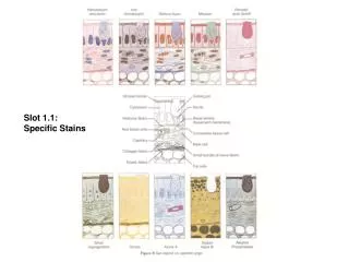
SPECIAL STAINS Drhuzlindahussin Dept of pathology 13 march 2018
H&Estainisroutinestain. • It isthepreliminary orthefirststainappliedtothetissue • sections • Givesdiagnosticinformationinmostcases. • Aspecialstainisastainingtechniquetohighlight various individual tissue component once we have preliminaryinformationfromtheH&Estain
CLASSIFICATION: Stains forcarbohydrates Stains foramyloid Nucleic acidstains Lipidstains Stains formicroorganisms Connective tissuestains Stains for pigments andminerals
CARBOHYDRATES SIMPLECARBOHYDRATES (moleculescomposedpurelyof carbohydrates) GLYCOCONJUGATES (molecules composed of carbohydrates and other molecules suchas protein andlipid) • Monosaccharides (glucose,mannose,galactose) • Oligosaccharides (sucrose,maltose) • Polysaccharides • (glycogen,starch) • Proteoglycans • Mucins • Othersglycoproteins
1. STAINS FOR CARBOHYDRATE • Periodicacidschiff(PAS)technique • Alcianbluemethod • Combined alcianblue-PAS techhnique • Mucicarminetechnique • Colloidal irontechnique • Metachromaticstaining • Iodine staining forglycogen • Enzymatic digestiontechnique Diastase digestion, Sialidase digestion, Hyaluronidasedigestion
Candida in lung, PAS stain. Glycogen in Ewing's sarcoma, PAS stain This is nodular glomerulosclerosis involving a glomerulus in the kidney, as seen with the PAS stain.
Note PAS +ve cytoplasmic granules partly resistant to diastase
Alcian blue/ PAS stain Acid mucins - blue. Neutral mucins - magenta. Mixtures - purple. Nuclei - grey/blue. The goblet cells of the gastrointestinal tract are filled with abundant acid mucin and stain pale blue with this Alcian blue stain.
CRYPTOCOCCUS STAINED BY MUCICARMINE The cytoplasm of the cells lining this neoplastic gland in a colonic adenocarcinoma are pink with the mucicarmine stain, indicative of mucin production typical for an adenocarcinoma at this site
2. STAIN FOR AMYLOIDI. Congo red -Also called amyloid stain -Must examine stained tissue with standard and polarized light -Amyloid under polarized light has apple green birefringence, based on the molecule being in an antiparallel beta-pleated sheet. - amyloid-red; nuclei- blue
AMYLOID • Extracellular,amorphous,eosinophilic material • Composedofproteininanantiparallel- pleated sheetconfiguration • InH&Estain,canbeconfusedwithhyaline and fibrinoidsubstances • Earliestspecialstainusedforamyloidwas Iodine byVirchow
Amyloid displays apple-green birefringence under polarized light. AMYLOID BY CONGORED STAIN UNDER LIGHT MICROSCOPE
3. Stains for lipid • Oil Red O • Sudan black B
LIPIDS SIMPLELIPIDS COMPOUND LIPIDS DERIVEDLIPIDS • FATS • OILS • WAXES • Derived from simple or compoundlipids byhydrolysis • cholesterol • Bileacids - c/o fatty acids, alcohol and one moregroupsuchas phosphorus or nitrogen
Simplelipidisbestdemonstratedwithfreshfrozen sections • Bestfixative–Formalcalcium(2%calciumacetate+ 10%formalin)
I. Oil Red O • Stain identifies neutral lipids and fatty acids • Fresh smears / cryostat sections of tissue are necessary because alcohols used in tissue processing remove lipids • Rapid and simple routine stain • Uses: • Differentiate fibroma from thecoma (not that important a distinction) • Diagnose renal cell carcinoma, sebaceous gland tumors of skin, lipid-rich carcinomas • Identify fat emboli in lung tissue or clot sections of peripheral blood