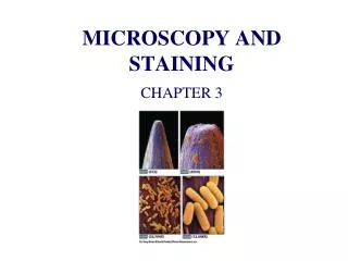
Microbial Staining Techniques
140 likes | 258 Vues
Learn about the importance of stains in microbiology, staining principles, different types of dyes, and staining techniques like Gram and endospore staining. Understand how dyes bind to cells and reveal microbial structures.

Microbial Staining Techniques
E N D
Presentation Transcript
Stains and objectives of staining • Stains are useful for the following reasons • It makes the microscopic semi-transparent objects visible • To study the shape and size • To reveal the presence of various internal and external structures • To produce specific chemical and physical reaction • A colouring agent that is used fro general purposes is called a dye
The one that is used for biological purposes is called a stain • acidic, basic and neutral • An acid (or anionic) dye has a negative change. Eg., Eosin, Rose Bengal, Acid fuchsin • Basic dye (or cationic) carries a positive charge. eg., Methylene Blue, basic fuchsin, crystal violet, malachite green, safranin • Basic dyes bind to negatively charged molecules like nucleic acid and many proteins • Bacterial cells surfaces are negatively charged • Neutral dye is a complex salt of a dye acid with a dye base
Dye Characteristics • Dyes have two features in common • chromophoregroups bind with cells by ionic, covalent or hydrophobic bonding • Positive staining procedure, a positively charged, chromophoreis attracted to the negatively charged outer surface Methylene blue • Negative staining procedures, negatively charged chromophorerepelled by the negatively charged microorganisms • Nigrosin and Indian ink
Staining reactions • When the pH of the surroundings of the microbial cells is either neutral or alkaline, all microbial cells have a negative charge on their surface, called the surface charge • microbes from acidic environments stain poorly with basic dyes • basic dyes are made up as alkaline solutions • potassium hydroxide (OH) is added to solutions of methylene blue Loeffler’s Methylene blue
Some bacteria excrete alkaline materials during growth • cell surface has a greater negative charge • Basic dyes stain microorganisms better under neutral or alkaline conditions • If the dye base molecule has a negative charge, it is repelled by the cell’s negatively charged surface • negatively charged dyes neither bind to the cell’s surface nor are they able to penetrate into the cell • acid dyes • Negatively charged dyes are called negative stains
Simple staining • simple staining solution, contains only one stain • microorganisms give the colour characteristic of the staining solution • purpose of simple staining to reveal the size and shape • simple stains, carbol fuchsin, crystal violet, methylene blue
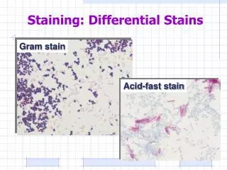
Differential Staining • more than one dye is employed • Differential staining divide the bacteria into separate groups • Gram stain and Acid-fast stain
Gram Staining • In 1884 • Christian Gram discovered a new technique to differentiate the bacteria of similar morphology
Principle • Gram – positive bacteria will retain the crystal violet and appear deep violet in colour • Gram-negative bacteria lose the crystal violet on decolorization are counter stained by the safranine, appear red in colour • reactions are associated with the structure and composition of the cell wall • Gram-negative bacteria are thinner, contain a higher percentage of lipid content • alcohol treatment extracts the lipid
results in increased porosity or permeability of the cell wall • violet-iodine complex, can be extracted, decolorized • take up the colour of the counter stain safranin • Gram-positive bacteria lower lipid content dehydrated • pore size decreased, permeability is reduced, CV-I complex can - not be extracted
Endospore Staining • Endospores strongly resist application of simple dyes, but once stained are quiet resistant to decolorization • simple stains are used, the body of the bacillus is deeply colored, whereas the spore is unstained and appears as a clear area in the organism • By vigorous staining procedures the dye can be introduced into substance of the spore • contrasting counter stain is usually applied in the ordinary fashion initial stain taken up by the spore, second stain appear in the cytoplasm