Gram Staining
Gram Staining. Differential Stain. Differential Stain Distinguishes between different groups of bacteria based on their staining ability. How can you tell a bacterial cell from normal body cells when a possible infection has taken place?.


Gram Staining
E N D
Presentation Transcript
Gram Staining Differential Stain
Differential Stain • Distinguishes between different groups of bacteria based on their staining ability
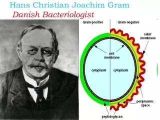
How can you tell a bacterial cell from normal body cells when a possible infection has taken place? Problem presented to Hans Christian Gram (1884) while studying origins and causes (Ethiology) of pneumonia. Couldn’t tell lung tissue from Streptococcus pneumoniae. Developed Gram staining technique which is unique to bacterial cells due to their cell wall construction
How it works • Gram positive bacteria have a thick layer ofPeptidoglycan(up to 90%)in their cell wall • Gram negative bacteria have a thinner layer (5- 20%)of this glycoprotein. • **Most cells are Gram -, except for Gram + bacteria, yeast and a few molds Two derivatives of glucose N-acetylglucosamine (NAG) N-acetlymuramic acid (NAM).
Gram PositiveGram Negative Peptidoglycan Porin Lipids + LSP Bilipid
How to do it • To a properly prepared, heat fixed smear, add 1 drop of the PRIMARY STAIN – Crystal Violet. Wait 1 minute and then rinse. • Add the MORDANT (intensifier) – Gram’sIodine. Wait 1 minute and rinse • DECOLORIZEwith Alcohol. Alcohol will remove stain from thin layers of peptidoglycan. Gram + hang onto stain while Gram – stain is washed away. 1 second, rinse then 3 – 5 seconds and rinse SECONDARY STAIN or COUNTERSTAIN is then applied give decolorized stain a stain. Use Safranin. Wait 1 minute and rinse
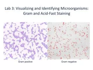
The results • Gram Positive stains get colored by crystal violet and safranin so they will be purple. • Gram Negatives will only be stained now with Safranin and be pink.
Problems with the procedure? • Leaving the alcohol on too long. Will decolorize even Gram + cells • Too much of a rinse after the Crystal violet. Washes away all the stain • Not enough Mordant (Iodine) to intensify primary stain. • Old cultures. Cells break down due to not enough nutrients present.
Bacteria for lab • Bacillus subtilis • Aquaspirillum serpens • Branhamella catarrhalis • Sarcina aurantiaca • Serratia marcescans