Staining Microorganisms
Staining Microorganisms. An overview of staining. Staining. Coloring organism with a dye Microorganisms must be FIXED to microscopic slide first Kills microbe Attaches organism to the slide Preserves various parts with minimal distortion Steps to fix:


Staining Microorganisms
E N D
Presentation Transcript
Staining Microorganisms An overview of staining
Staining Coloring organism with a dye Microorganisms must be FIXED to microscopic slide first • Kills microbe • Attaches organism to the slide • Preserves various parts with minimal distortion Steps to fix: • SMEAR is placed on slide and allowed to dry • Passed through Bunsen burner several times • Stain then applied and washed off
STAINING • Stains are salts composed of positive and negative ions (one is colored and is called a chromophore) Basic dyes-color is positive ion Acidic dyes- color is negative ion Bacteria are slightly negatively charged at pH 7.0, so basic dye is attracted more to them. Includes: crystal violet methylene blue malachite green safranin
STAINING • Acidic dyes not attracted to bacteria because stain’s negative ion is repelled • Used to stain background instead (some bacteria are colorless). This is called negative staining • Great when fixing is not possible because only background is stained
STAINING TYPES OF STAINING: • Simple • Differential • Gram Stain • Acid Fast • Special • Negative staining for capsules • Endospore Staining • Flagella Staining
Staining • Simple Sometimes an additive called a mordant. It increases uptake of the dye.
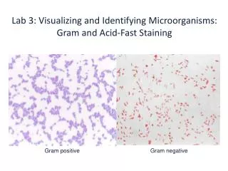
Staining • Differential • Gram Stain (developed in 1884) Procedure: 1. apply heat. this fixes smear; slide is covered with purple dye (primary stain) 2. dye is washed off and the mordant, iodine, is placed and washed off 3. slide washed with alcohol (decoloring agent) 4. slide is re-stained with safranin(red dye is a counterstain)
Staining • Differential • Acid Fast • Binds strongly to waxy material in cell walls Procedure: 1. red dye carbolfuchsin is applied to a fixed smear and slide is heated 2. slide is cooled and washed with water and then acid-alcohol 3. If colorless, slide is counterstained with methylene blue
Staining • Special Stains • Negative Staining for capsules • Organism is placed in solution with India ink or nigrosin and then contrastained with safranin
staining • Endospore (spore staining) • Most common stain is Schaeffer-Fulton endospore stain • Malachite green (primary stain) is applied to heat-fixed smear and heated to steaming for 5 min • Washed with water • Next safranin (counterstain)
Staining • Flagella Staining • Uses a mordant and carbolfuchisin