Download
1 / 29
300 likes | 389 Vues
Explore the fascinating world of neuroanatomy and neurotransmitters to understand how our biology influences our behavior and mental processes. Learn about neurons, neurotransmitters, synaptic firing, and the functions of the central and peripheral nervous systems.

E N D
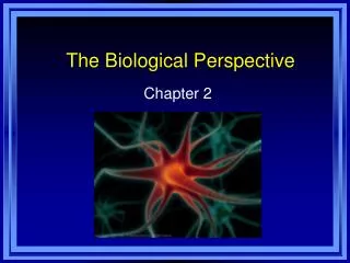
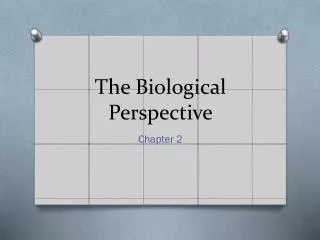
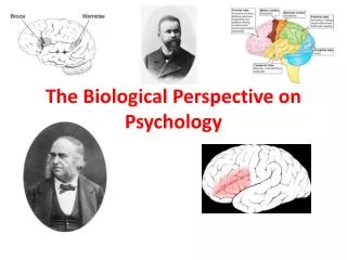
Chapter Two - Pearson R. M. TOLLES It’s ALIVE!!!!! PsychologyThe Biological Perspective
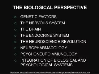
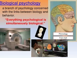
The Beginning – Module 4Explain how messages are transmitted by the neurons, and describe the functions of the spinal cord and the peripheral nervous system. • Everything psychological is a simultaneously biological. • Franz Gall did not subject his beliefs about phrenology to scientific test, but this early theory did help scientists to begin thinking about links amount our biology, behavior, and mental processes.
Neuroanatomy refers to the study of parts and functions of neurons. Parts of a neuron: Dendrites – root-like parts of the cell that stretch out form the cell body. Cell body (also called the soma) – contains the nucleus and other parts of the cell needed to sustain its life Axon – wire-like structure ending in the terminal buttons that extend form the cell body Myelin sheath – a fatty covering around the axon of some neurons that needs neural impulses Terminal buttons (also called end buttons, terminal branches and synaptic knobes) – the branched end of the axon that contains neurotransmitters Neurotransmitters – chemical contained in terminal buttons that enable neurons to communicate. Neurotransmitters fir into receptor cites on the dendrites or neurons like a key fits into a lock Synapse – the space between the terminal buttons of one neuron and the dendrites of the next neuron. Neuroanatomy
How a Neuron “Fires” • Neural firing is an electrochemical process. • Electricity travels within the cell (from the dendrites to the terminal buttons), and chemicals (neurotransmitters) travel between cells in the synapse. • Electricity does not jump between the neurons • The electric message firing is called an action potential. • When a neuron fires completely or it does not fire at all, this is called the all-or-none principle. The impulse is the same size every time.
Neurotransmitters • Different types of neurotransmitters exist • Types: • Excitatory – they excite the next cell into firing • Inhibitory – they inhibit the next cell from firing • Each synaptic gap at any time may contain many different kinds of inhibitory and excitatory neurotransmitters. • The amount determine whether it will pass the threshold and fire
How Neurotransmitters Influence Us • Each neurotransmitter travels a designated path in the brain and has a particular effect on behavior and emotions. • Acetylcholine, one of the best-understood neurotransmitters, affects muscle actions, learning, and memory. • The endorphins are natural opiates released in response to pain and exercise
The Nervous System • One major division of the nervous system is the central nervous system (CNS), which consists of the brain and spinal cord. • The other is the peripheral nervous system (PNS), which consists of the neurons that connect the CNS to the rest of the body by means of nerves (bundles axons of the sensory and motor neurons) • Sensory neurons carry incoming information from sense receptors to the CNS
Nervous System • Interneurons communicate within the CNS and between sensory and motor neurons. • Motor neurons carry information from the CNS out to the muscles and glands. (also called Efferent Neurons) • Organization of the nervous system: • Divided into different catergories based on function.
SECTION 1: Explain how messages are transmitted by the neurons, and describe the functions of the spinal cord and the peripheral nervous system. Nervous System: (WEB)
The Central Nervous System • Reflex pathways are automatic inborn responses to stimuli, and they do not rely on conscious decisions made in the brain. • A signal sensory neuron, excited by some stimulus (such as flame), passes a message to an interneuron in the spinal cord. • The interneuron activates a motor neuron, causing some muscle reaction (such as jerking away). • In contrast, neural networks found in the brain are clusters of many neurons that together share some special task. • These complex networks strengthen with use, learning from experiences. • Each neural network connects with other networks performing different tasks.
The Peripheral Nervous System Peripheral Nervous System (PNS) has two main divisions. • The Somatic Nervous System enables voluntary control of the skeletal muscles. • The Autonomic Nervous System, through its sympathetic and parasympathetic divisions, controls our involuntary muscles and glands
Module 5: Identify the major structures of the brain, and explain the functions of each structure.
The Tools of Discovery • Clinical observations have long revealed the general effects of damage to various areas of the brain. But MRI scans now reveal brain structures and EEG, PET, and fMRI (functional MRI) recordings reveal brain activity. • By surgically lesioning or electrically stimulating specific brain areas, by recording the brains surface electrical activity, and by displaying neural activity with computer-aided brain scans, neuroscientists explore the connections among brain, mind, and behavior.
Ways of Studying the Brain • Accidents • Lesions • Electroencephalogram (EEG) • Computerized Axial Tomography (CAT or CT) • Magnetic Resonance Imaging (MRI) • Positron Emission Tomography (PET) • Functional MRI • Phineas Gage
Older Brain Structures • The brainstem is the oldest part of the brain and is responsible for automatic survival functions. • Its components are: • The medulla (which controls heartbeats and breathing) • The pons (which helps coordinate movements) • The reticular formation (which affects arousal). • The thalamus, the brain’s sensory switchboard, sits above the brainstem. • The cerebellum, attached to the rear of the brainstem. coordinates muscle movement and helps process sensory information.
The Limbic System • Between the brainstem and cerebral cortex is the limbic system, which is linked to emotions, memory, and drives. • One of its neural centers, the amygdala, is involved in responses of aggression and fear. • Another, the hypothalamus, is involved in various bodily maintenance functions, pleasurable rewards, and the control of the hormonal system. • The hypothalamus sits just above the pituitary (the “master gland”) and controls it by stimulating it to trigger the releases of hormones. • The hippocampus also part of the limbic system, processes memory.
The Cerebral Cortex • The cerebral cortex is the thin surface layer of interconnected neurons covering the brain’s hemisphere. • The human brain’s cortex is larger than that of other animals, and it enables learning, thinking, and the other complex forms of information processing that make us uniquely human
The Structure of the Cortex • Each cerebral hemisphere has for geographic areas. • Frontal lobe - just behind the forehead, is involved in speaking, muscle movement, and planning and judgment. • Parietal lobes - top of the head and toward the rear, receive sensory input for touch and body position. • Occipital lobes - back of the head, include visual areas. • Temporal lobes - just above the ears, include auditory areas. • Each lobe performs many functions and interacts with other areas of the cortex
Functions of the Cortex. • Some areas of the brain serve specific functions. • Motor cortex - an arch shaped region at the rear of the frontal lobes, controls voluntary movements. • Sensory cortex - a region at the front of the parietal lobes, registers and processes body sensations. • In these regions, body parts require precise control (in the motor cortex) or those that are especially sensitive (in the sensory cortex) occupy the greatest amount of space. • Most of the brain’s cortex – the major portions of the four lobes – is devoted to uncommitted association areas, which integrate information involved in learning, remembering, thinking, and other higher-level functions.
Language • Language results from the integrations of many specific neural networks performing specialized subtasks. • When you read aloud, your brain’s visual cortex registers words as visual stimuli, the angular gyrus transforms those visual representations into auditory codes, • Wernicke’s area interprets those codes and sends the message to Broca’s area which controls the motor cortex as it creates the pronounced words.
The Brain’s Plasticity • If one hemisphere is damaged early in life, the other will pick up many of its functions. • This plasticity diminishes later in life, although nearby neurons may partially compensate for damaged ones after a stroke or other brain injury.
Our Divided Brain • Clinical observation long ago revealed that the left cerebral hemisphere is crucial for language. • Split-brain research (experiments on people with a severed corpus callosum) has confirmed that in most people the left hemisphere excels in visual perception and the recognition of emotion. • Studies of healthy people with intact brains confirm that each hemisphere makes unique contributions to the integrated functioning of the brain.
Brain Organization and Handedness • About 10% of us are left-handed. • Almost all right-handers process speech in the left hemisphere, as do more than half of all left-handers. • The remainder of left-handers split about evenly in processing language in the right hemisphere of in both hemispheres. • The percentage of lefties decreases sharply with age, from about 15% at age 10 to less than 1% at age 80. • This decline reflects a higher risk of accidents.
Section 3: Identify the hormones secreted by the major glands of the endocrine system and the role each plays. • Endocrine System
The Endocrine System • The endocrine system is a set of glands that secret hormones into the bloodstream. • These chemical messengers travel through the body and affect other tissues, including the brain. • Some hormones are chemically identical to neurotransmitters. • The endocrine systems master gland, the pituitary influences hormone release by other glands. • In an intricate feedback system the brain’s hypothalamus influences the pituitary gland which influences other glands which release hormones which in turn influence the brain.
Module 6: Explain the role of chromosomes and genes in heredity, and evaluate the methods used by psychologists to study the role of heredity in determining traits Heredity and Genes Define - inherit form parents/map of characteristics Physical and Psychological traits are passed Genes and Chromosomes Genes - building blocks of heredity Genes are in chromosomes
Section 4: Continued • Web on Genes and Chromosomes
Section 4: Continued… Nature vs Nurture What is Nature vs Nurture? Kinship Studies Twin studies Adoptee Studies Twins reared apart