Download
1 / 38
380 likes | 850 Vues
Giardia Lamblia. Artistic impression by Russel Kightley Source: http://soils.cses.vt.edu. Giardia. Giardia lamblia is a flagellated protozoan that infects the duodenum and small intestine . R ange from asymptomatic colonization to acute or chronic diarrhea and malabsorption.

E N D
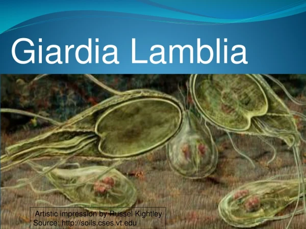
Giardia Lamblia Artistic impression by Russel Kightley Source: http://soils.cses.vt.edu
Giardia • Giardia lambliais a flagellated protozoan that infects the duodenum and small intestine. • Range from asymptomatic colonization to acute or chronic diarrhea and malabsorption. • More prevalent in children
EPIDEMIOLOGY • Geographical distribution-world wide • Usually occurs sporadically • Major reservoir for spread :water contaminated with giardia cysts • Giardia cysts are relatively resistant to chlorination and to ultraviolet light irradiation • Boiling is effective for inactivating cysts.
Transmission F:eco-oral • Habitat - Duodinum and upper part of ileum of man
morphology - Exists in two phases-trophozoite and cyst form Trophozoite form-when viewed flat-appears like tennis or badminton racket-when viewed side –on-resembles –spit pear Size-14µm long Anterior end is broad and rounded-posterior end tapers Is bilaterally symmetrical & all organs are paired
Tear drop shaped • 2 adhesive discs, • 2 median bodies, • 2 nuclei • 4 pairs of flagella
CYST–is fully formed cyst –oval in shape =12µm long 7µm broad The axostyles lie –more or less diagonally-form sort of dividing line There are four nuclei –remain clustered at one end An acid environment-often cause parasite to encyst
four pairs flagellae a flat ventral surface sucking or adhesive disk 8 to 12 mm long and 7 to 10 mm wide convex dorsal surface Source: http://soils.cses.vt.edu
LIFE CYCLE life cycle of G. lamblia is composed of 2 stages: • trophozoites • cysts
In tropozoite stage Parasite multiplies(intestine of man –binary fission) during unfavourable condition in duodenum(encystment occur during encystment cells then divide into two within the cyst infection to man is brought by ingestion of cyst
Trophozoites : Lives in duodenum, jejenum and upper ileum They come in close contact to the mucosal, but do not invade the host. Adhesive disc fits over surface of epithelial cell The flagella act as a pump to move nutrients away from the microvilla and hold the adhesive disc near the mucosa. Rapid division to produce large numbers quickly
Pathogenesis and Immune response • The production of diarrhea, and occasionally malabsorption, is the result of a complex interaction of Giardia with the host, • Infection occurs after oral ingestion of as few as 10 to 25 cysts. • After excystation, trophozoites colonize and multiply in the upper small bowel • Adherence of G. lamblia in the human gut may be via the disc • but may also involve specific receptor-ligand interactions
Pathogenesis and Immune response Several pathogenic mechanisms have been postulated Disruption of the brush border Mucosal invasion Elaboration of an enterotoxin Stimulation of an inflammatory infiltration leading to fluid and electrolyte secretion and occasionally to villous changes
Ventral sucking disc Source: Gallery of histology Woods and Ellis2000 TEM micrograph showing the method of attachment to the duodenal wall.
Immune Response Partially protective immunity may develop to Giardia Immune response involves both cellular and humoral immunity Ig A, serum Ig G and Ig M are detected in patients: role of Ig A is not completely understood, probably inhibits trophozoite attachment IgA deficiency lead to chronic giardiasis Cell mediated immune response may also play a role Human milk may also play a role in protection of the host against Giardia : Free fatty acids and IgA antibodies
CLINICAL MANIFESTATIONS • incubation period :1–2 wk • clinical manifestations :asymptomatic . acute infectious diarrhea, chronic diarrhea with failure to thrive and abdominal pain or cramping. • Symptomatic infections occur more frequently in children than in adults. • Most symptomatic patients : acute diarrhea. low-grade fever, nausea, and anorexia; • intermittent or more protracted course characterized by diarrhea, abdominal distention and cramps, bloating, malaise, flatulence, nausea, anorexia, and weight loss develops
CLINICAL MANIFESTATIONS • stools may be profuse and watery and later become greasy and foul smelling • Stools do not contain blood, mucus, or fecal leukocytes • Varying degrees of malabsorption may occur.
Abnormal stool patterns may alternate with periods of constipation and normal bowel movements. • Malabsorption of sugars, fats, and fat-soluble vitamins has been well documented and may be responsible for substantial weight loss. • Giardiasis has been associated with growth stunting and repeated Giardia infections with a decrease in cognitive function in children in endemic areas.
Giardiasis should be considered in young children in child care or in any person who has had contact with an index case or a history of recent travel to an endemic area
DIAGNOSIS • Stool examination: • Microscopic examination for trophozoites or cysts • Stool enzyme immunoassay (EIA) or direct fluorescent antibody tests are more sensitive • Aspiration or biopsy of the duodenum or upper jejunum -Enterotest
Enterotest Uses a coiled thread inside small weighted gelatin capsule Swallowed after attaching the free end of the thread to cheek Capsule passes through stomach to duodenum After 2 hours, thread is withdrawn ,placed in saline Centrifuged deposit of saline is examined for giardia
TREATMENT • should receive therapy : acute diarrhea failure to thrive exhibit malabsorption
Treatement • Metronidazole (250mg TID -5 days) • Trimidazole(2gms OD) • Furozolidone(100mg QID)-7-10 -days
PREVENTION • Handwashing • purify public water supplies adequately include chlorination and filtration. • Travelers to endemic areas are advised to avoid uncooked foods that might have been grown, washed, or prepared with water that was potentially contaminated. • Purification of drinking water can be achieved by a filter or by brisk boiling of water for at least 1 min
Genus Trichomonas • Its includes a group of flagellated protozoa It infect humans and animal • 3 species of trichomonads found in human. • Two are normally harmless. • T. vaginalis which is a serious sexually transmitted pathogen.
Trichomonas vaginalis: • It is the etiological agent of trichomoniasis. • Trichomoniasis is a common sexually transmitted disease with a worldwide distribution. • Transmittable, sexually and through contact with toilet seats and towel. • T. Vaginalis despite it name, infect both men and women. • In females the organism inhabits the vagina and urethra • In males it is found in the urethra, prostate or, seminal vesicles.
The life cycle consist only of a trophozoite stage which is transmitted by direct contact during sexual intercourse
T. Vaginalis trophozoite • Fg=flagella • Bb=basal body • Nu=nucleus • Ax=axostyle • um=undulating membrane • Cy=cytostomal groove • Cs=costa
(B) T. vaginalis on the surface of a vaginal epithelial cell prior to ameboid transformation. 5 μm
(C) Ameboid morphology of T. vaginalis as seen in cell culture. 5 μm
Life cycle:flagella are found in the genital tract. Transmission: during dealings with contaminated matters (clothes, cotton..etc.
Sign and symptoms: • T. vaginalis causes different manifestation in men and women. • Women are more likely to exhibit symptoms which tend to persist longer • Incubation period is 4-28 days. • In female: ranges from asymptomatic, to mild or moderate irritation, to extreme vaginitis • 10 - 50%: asymptomatic • The commonest symptoms: vaginal discharge, vulval itching, dysuria, or offensive odor , rare abdominal discomfort • Extreme cases associated with vulvitis and vaginitis • 2%: strawberry cervix appearance to the naked eye. • In male:50-90% are asymptomatic • mild dysuria or pruritus • minor urethral discharge
Vaginitis - Trichomoniasis • Profuse, frothy discharge, yellow-greenish in color foul odor, vulvar pruritus • Patchy vaginal erythema and (strawberry cervix)
Diagnosis: 1-Specimens: • vaginal discharge • urine sediment • prostatic secretion 1.Vaginal pH 2. Whiff test 3.Wet mount 4. Pap smear 5. Culture 6. Direct immunoflouresence assay 7. Polymerase chain reaction 8. Evaluation for other STDs
Upon application of 10% potassium hydroxide (KOH) to a vaginal swab sample, a fishy odor is released, which can suggest trichomoniasis or bacterial vaginitis. Potassium hydroxide amine test (Whiff test) : Whiff test: 10 % KOH