Download
1 / 1
10 likes | 270 Vues
Isolation and Identification of Chroococcidiopsis Photosystems Brian Moran 1 , Stephen Richardson 2 , Li Meng 3 , Brendan Williams 3 , Barry D. Bruce 3

E N D
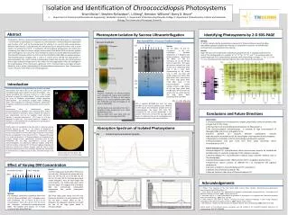
Isolation and Identification of Chroococcidiopsis Photosystems Brian Moran1, Stephen Richardson2, Li Meng3, Brendan Williams3, Barry D. Bruce3 Department of Chemical and Biomolecular Engineering, Vanderbilt University; 2. Department of Biochemistry,Maryville College; 3. Department of Biochemistry, Cellular and Molecular Biology, The University of Tennessee, Knoxville T. Elongatus 3A) 5A) TE Ch TE Ch kDa 1 2 3 4 5 6 7 1068 1068 700 e 700 356 356 g b h i Chroococcidiopsis 3B) c k m d f j a l 3D) Ch 10% 20% 30% Abstract Photosystem Isolation By Sucrose Ultracentrifugation Identifying Photosystems by 2-D SDS-PAGE DM% .6 .8 1 2 3 4 .6 .8 1 2 3 4 • Photosystem I (PS I) is a protein complex that transfers electrons from plastocyanin or cytochrome c to ferredoxin in cyanobacteria in response to photo-excitation. Understanding the organization and structure of PS I will lay the ground work for utilizing it as a part of solar energy devices. Different from plant PS I, cyanobacterial PS I was believed to be exclusively trimer, until a recent report of a tetramer form of PS I in Anabaena.Chroococcidiopsis photosystems are studied here considering their vitality in various extreme environments. Blue-Native PAGE and sucrose gradient ultra-centrifugation are used as the main methods to analyze and isolate different photosystems. The isolated Photosystems from the cells are further identified by SDS-PAGE and spectral analyses. Thermosynechococcus elongatus (TE) is used as a control and to identify the photosystems in Chroococcidiopsis. Our results indicate a photosystem complex that may be a PS I tetramer/trimer with a higher molecular weight than TE PS I trimer. This new organization of PS I will shed light on the understanding of the evolutionary significance of PS I monomerization and oligomerization. Hopefully, with a better understanding of Chroococcidiopsisphotosystems, these Photosystems can be utilized to create better solar energy apparatus. Blue NativePAGE of Sucrose Gradient Samples Sucrose Gradients Method: To further characterize the photosystem components of Chroococcidiopsis, bands from Blue NativePAGE separation (Fig 5A) were excised and subjected to separation via SDS-PAGE Gel electrophoresis and subsequent silver staining. 3C) Results: 3C) The labels, TE and Ch, correspond to the non-centrifuged thylakoid membranes of T. Elongatus and Chroococcidiopsis, respectively. The labels, 1-2 and 3-7, correspond to 3A and 3B (the same in Figure 4A). The upper gel has been stained. Lane 1 has no PSI trimer (1068 kDa) while lane 2 has a dark band corresponding to PSI trimer. Lane 3 does not have the middle large green band (~890 kDa) while 6 and 7 have little of the lowest large green band (~420 kDa). None of the Chroococcidiopsissucrose samples contain a high degree of the top large green band (~1180 kDa). This contrasts the results from 2A, which had the top large green band (solubilized with 1.0% DM). Results: Figure 5B shows the sample protein separations compared to the T. elongatus purified trimer control. The banding patterns in samples “a,” “e,” “f,” “g,” “h,” “k,” and “l” are consistent with the protein fragments in PS I (noted by blue asterisks), while the banding in samples “c,” “d,” “i,” j,” and “m” are consistent with the expected fragment sizes of PS II (noted by red asterisks). * * * Introduction * * * 1A As the consumption of energy continues to increase worldwide (now greater than 425 X 1018 J), the demand for novel and renewable energy capture devises has never been higher. One rapidly developing area of energy research is in solar energy utilization and bioelectric hybrid systems. These systems involve the union of biological photosynthetic protein complexes (photosystems) and electrically charged metal scaffolding, which, when organized in a particular orientation, allows them to together create the movement of electrons and thus electric current. Method: Thylakoid membranes of Chroococcidiopsis and T. elongatus (0.2 ug/uL) were solubilized with 1% DM for 1.5 hours at 25°C before loading on to sucrose gradients (10%--30%) with 0.05% DM and ultra-centrifuging for 16 hours at 20000 RPM . 3A) Two bands (1-2) were taken from the T. elongatus sucrose gradient to run on a Blue NativePAGE gel (3C). 3B) Five bands (3-7) were taken from the Chroococcidiopsis sucrose gradient (3C). 1A 3D) A separate BN-PAGE gel tests the same variables as the sucrose gradient prep (Method on left) except the membrane samples were not ultracentrifuged. The labels, 10%, 20%, 30%, correspond to the sucrose percentage in the sample. These lanes all have the top large green band. It is likely that the act of centrifugation and sedimentation results in the loss of this band. Photosystems from a cyanobacterial species, Thermosynechococcus elongates, have previously been studied for their potential in bioelectric devices because the species is well characterized and has an available crystal structure. Typically, T. elongates photosystem I (PS I) exists as a trimer (depicted in Figure 1A). Until recent report of a tetramer in Anabaena, it was believed that all cyanobacterial photosystems existed exclusively as trimers. Conclusions and Future Directions Conclusions: 1.Chroococcidiopsis has a Photosystem I complex, potentially a trimer or tetramer, that is larger than TE PS I trimer; 2.The majority of Chroococcidiopsis photosystems are PhotosystemI; 3.The Chroococcidiopsis trimer/tetramer is sensitive to high concentrations of detergent (DM) and ultra-centrifugation force; 4.Sucrose gradient can separate different photosystem mixtures with phycocyanin attached but not for intact higher order tetramer/trimer complexes. 5.Phycocyanin is attachedto the Chroococcidiopsis photosystem samples. 6.Chroococcidiopsis may have more than three states (monomer, dimer, trimer/tetramer) of PS I. Future directions and study: Chroococcidiopsis PS I trimer/tetramer, dimer and monomer need to be isolated and purified further to study the properties of the molecular complex. 1.ChroococcidiopsisPS I trimer/tetramer isolation using moderate methods such as Chromatography; 2.Componental analysis of the different forms of PS I using Mass Spectrometry; 3.Fluorescence spectra analysis of different PS I to characterize the pigment components; 4.Dynamic equilibrium assay of phycocyanin PS I association; 5.Stability assay of Chroococcidiopsis PS I; 6.Electron transport rates assay of Chroococcidiopsis PS I. 1B Absorption Spectrum of Isolated Photosystems Another genus of cyanobacteria that may exist as a tetramer and has recently become of interest in the science of bioelectrics is Chroococcidiopsis (Fig 1B). This organism is of particular interest because of its ability to survive a wide range of extreme environments that are uninhabitable to most other life forms. The goal of this study is to isolate photosystem I of Chroococcidiopsis and characterize the complex through various experimental techniques such as Blue NativePAGE and SDS Page electrophoresis, sucrose gradient ultracentrifugation, and Absorbance spectrum analysis. Using these methods to better understand PS I of Chroococcidiopsis, hopefully will determine whether this protein complex could be used in future bioelectric device research and, ultimately, expedite the introduction of similar devices as viable sources of clean, renewable energy in the future. 4A) Isolated Sucrose Gradient Samples 1A)T.E. Trimer with PsaL shown. 1B) Chroococcidiopsis microscopic image Effect of Varying DM Concentration Result: 2A) Three large green bands (PSI or PSII) and at least six other faint bands were separated (ref. Fig 7) for the 0.6, 0.8, and 1.0% DM. Yet, for the 2.0, 3.0, and 4.0% the top large green band mostly disappears. Also, for those higher DM concentrations, the middle large green band becomes larger. This suggests that with higher DM, the multicomplex photosystem disassociates into smaller photosystem complexes. 2B) Three large green bands and one blue band were detected. The concentration of DM did not have a drastic effect on the T. Elongatus. The large green bands do not match the size of the large green bands in Chroococcidiopsis. 2A) Chroococcidiopsis 2B) T. elongatus 4B) PSI Trimer 4C) Phycocyanin Acknowledgements 1. Phillips, Tony. Greening of the Red Planet. NASA Science News. [Online]. http://science.nasa.gov/science-news/science-at-nasa/2001/ast26jan_1/ 2. Lavan, David and Jennifer N. Cha. Approaches for biological and biomimetic energy conversion. Proceedings of the National Academy of Sciences. 2006, 103(14): 5251-5255. 3. Watanabe, Mai et al. Novel Supercomplex Organization of Photosystem I in Anabaena and Cyanophora paradoxa. Plant and Cell Physiology. 2010, 52(1): 162-168. 4. Lewis, Nathan S and Daniel G. Nocera. Powering the planet: Chemical Challenges in Solar Energy Utilization. Proceedings of the National Academy of Science. 2006, 103(43): 15729- 15735. 5. Mai Watanabe , Masako Iwai, Rei Narikawa and Masahiko Ikeuchi. Plant Cell Physiology. 2009, 50(9): 1674–1680 Special thanks to the National Science Foundation and the funding of the TN-SCORE Research Experience for Undergraduates (NSF EPS-1004083). Method: The thylakoid membranes (prepared from fresh cells) of Chroococcidiopsis and T. elongatus were each solubilized for 1.5 hours at 25°C in six concentrations (0.6, 0.8, 1.0, 2.0, 3.0, 4.0 %) of the detergent n-dodecyl-β-ᴅ-maltopyranocide (DM). The samples were loaded on a 4-16% acrylamide Blue NativePAGE gel.