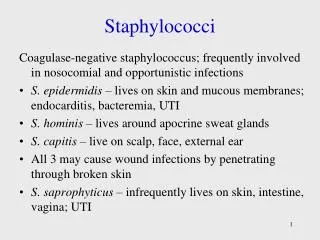
Isolation and Identification of Staphylococci
520 likes | 1.75k Vues
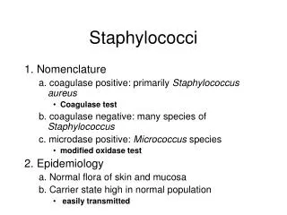
Isolation and Identification of Staphylococci. Gram Stain. Gram Positive coccus. Catalase. _. +. Streptococci. Oxidase. _. +. Staphylococci. Micrococci. Coagulase. _. +. Staphylococcus epidermidis. Staphylococcus aureus. CNS. Staphylococcus saprophyticus.

Isolation and Identification of Staphylococci
E N D
Presentation Transcript
Isolation and Identification of Staphylococci
Gram Stain • Gram Positive coccus Catalase _ + Streptococci Oxidase _ + Staphylococci Micrococci Coagulase _ + Staphylococcus epidermidis Staphylococcus aureus CNS Staphylococcus saprophyticus
Coagulase _ + Staphylococcus epidermidis Staphylococcus aureus CNS Staphylococcus saprophyticus Negative Positive Positive Negative Slide method Tube method Coagulase test
Non-hemolyticStaphylococcus species: Staphylococcus epidermidis
Staphylococcus saprophyticus: non-hemolytic, bright white, creamy colonies
Strains of Staphylococcus aureus produce a golden yellow pigment
Strains of Staphylococcus aureus not a golden yellow pigment producer
MANNITOL SALT AGAR ( MSA ) • INGREDIENTS • Peptone. • Beef Extract. • D-Mannitol .............. 1.0%. • Sodium Chloride ...... 7.5%. • Agar ......................... 1.5%. • Phenol Red. AS PH INDICATOR • Final pH 7.4 ± 0.2 at 25°C.
PRINCIPLEAND RESULTS • Mannitol Salt Agar is a nutritive medium due to its content of peptones and beef extract, which supply essential growth factors, such as nitrogen, carbon, sulfur and trace nutrients. • The 7.5% concentration of sodium chloride results in the inhibition of bacterial organisms other than staphylococci. • Mannitol fermentation, as indicated by a change in the phenol red indicator, aids in the differentiation of staphylococcal species.
Mannitol Salt agar Staph. epidermidis Staph. aureus
NB Staph saprophyticus Staph epidermidis Coagulase Negative Staph Novobiocin resistance Test: Procedure: 1. Inoculate blood agar plate with the test organism. 2. Aseptically apply Novobiocin disc onto the center of the streaked area. 3. Incubate the plate at 37oC for 24 hrs. 4. Accurately measure the diameter of the inhibition zone around the disc.
Novobiocin test A novobiocin disk will be placed on the plate, Novobiocin is an antibiotic that many Staphylococcus strains are sensitive to with the exception of one , Staph. saprophyticus thatis risist to novobiocin antibiotic.
Staphylococcus epidermidisGrowing on Blood Agar Note there is no hemolysis (gamma reaction) on the blood agar and the organism is sensitive to the antibiotic novobiocin as shown by the zone of inhibition.
Staphylococcus saprophyticusGrowing on Blood Agar Note there is no hemolysis (gamma reaction) on the blood agar and the organism is resistant to the antibiotic novobiocin.
Staphylococcus aureusGrowing on Blood Agar Note beta hemolysis (complete lysis of the red blood cells around the colonies; see arrows) on the blood agar and the organism is sensitive to the antibiotic novobiocin.
DNase TEST AGAR Media ingredients Enzymatic Digest of Casein.................1.5% Enzymatic Digest of Animal Tissue.....0.5% Sodium Chloride...................................0.5% Deoxyribonucleic Acid.............0.2% Agar........................................1.5% Final pH: 7.3 ± 0.2 at 25°C Principles of the Procedure The nitrogen, vitamin, and carbon sources are provided by Enzymatic Digest of Casein and Enzymatic Digest of Animal Tissue. Sodium Chloride provides essential ions while maintaining osmotic balance. Deoxyribonucleic Acid enables the detection of DNase that depolymerize DNA. Agar is the solidifying agent. Test Procedure 1. Inoculate plates by spotting or streaking a heavy inoculum of test organism. 2. Incubate plates at 35 ± 2°C for 18 – 24 hours and up to 48 hours. 3. Flood plates with 1 N HCl. 4. Observe for clearing around the spot or streak. Record results. Results A zone of clearing around the spot or streak indicates DNase activity.
Staphylococcus aureusGrowing on DNase Agar Note there is breakdown of the DNA in the agar. There is a clear zone (arrow) around the bacterial growth where there is no longer any DNA left in the agar to precipitate out of solution after the 1N HCL was added.
Staphylococcus epidermidisGrowing on DNase Agar Note there is no breakdown of the DNA in the agar. After adding the 1N HCl, the entire plate turned cloudy as the DNA precipitated out of solution. There is no clear zone around the bacterial growth.
DNase Test Agar with Methyl Green Methyl green forms a complex with intact (polymerized) DNA to form the green color of the medium. DNase activity depolymerizes the DNA, breaking down the methyl green-DNA complex, which results in the formation of colorless zones around colonies of the test organism. A negative test is indicated by the absence of a colorless zone around the colonies.
DNase Test Agar with Toluidine Blue Toluidine blue forms a complex with intact (polymerized) DNA. In the intact DNA complex, the toluidine blue has the normal blue color. Dnase activity depolymerizes the DNA, breaking down the dye-DNA complex. In the presence of nucleotides produced from the DNase depolymerization, the dye takes on its metachromatic color, forming pink to red zones around bacterial growth. A negative test is indicated when the medium remains blue.
Protein A Latex Test L Protein A is found on the cell surface of about 95 % of human strains of S. aureus and has the ability to bind the Fc portion of immunoglobulin G (IgG) L IgG S S L S S Fc L L S S=S.aureus with Protein A L=Latex particle S Protein A S
Staphyloslide™ Latex Test for Staphylococcus aureus Latex Test consists of latex particles coated with human fibrinogen and IgG. On mixing the latex reagent with colonies of staphylococci which have clumping factor or Protein A present, cross-linking will occur giving visible agglutination of the latex particles. Such agglutination will occur notably with S. aureus. If neither clumping factor nor Protein A are present, no agglutination will occur and the result will be regarded as negative.