Download
1 / 36
420 likes | 603 Vues
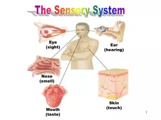
SENSORY SYSTEM. CHAPTER 13. Sensory System. Allows us to experience the world… see the trees hear voices of friends and family feel the heat of the sun taste favorite foods Allows us to keep track of what is happening within our bodies…. Receptors & Sensations. Cells that detect stimuli…

E N D
SENSORY SYSTEM • CHAPTER 13
Sensory System... • Allows us to experience the world… • see the trees • hear voices of friends and family • feel the heat of the sun • taste favorite foods • Allows us to keep track of what is happening within our bodies…
Receptors & Sensations • Cells that detect stimuli… • Receptor – is a specialized area of a sensory neuron that detects a specific stimulus • Example – eye responds to light • Sensory neuron transmit information to the CNS. • 5 Types of receptors • Pain receptors (nocioceptors) – stimulated by tissue damage or distention • Thermoreceptors – receptors stimulated by changes in temperature • Mechanoreceptors – receptors stimulated by changes in pressure or movement of body fluids • Photorecpetors – receptors stimulated by light
Sensation • Sensation is the conscious awareness of incoming information. “OUCH” – when you become aware of painful stimulus
4 Components to a sensation: • Stimulus • ex. light is a stimulus for sense of sight (in absence of light = no sight) • Receptor – light wave stimulates photoreceptor in the eye – produces a nerve impulse • Sensory nerve – nerve impulse is conducted by a sensory nerve to the occipital lobe of the brain • Special area of the brain • sensory information is interpreted as sight in the occipital lobe of the brain • Sensation is experienced by the brain and not by sensory receptors – sensory information has just stimulated a part of the brain.
Characteristics of Sensation:Projection & Adaption • If pain is experienced by the brain, why does my finger hurt? • Projection Process by which the brain, after receiving a sensation - refers that sensation back to its source. • “Phantom Limb” - example of projection, a patient may still feel pain in an amputated part. Severed nerve endings continue to send sensory information to the parietal lobe. The brain interprets the information as pain and projects the feeling back to the amputated area. This phenomena will decrease when the nerve endings begin to heal. • Phantom pain helps an amputee use artificial limbs (prosthesis). 6
Adaption when continuously stimulated, receptors send fewer signal tot he area of the brain that interprets sensory information • when you walk into a room that has a strong odor - at first it is overwhelming but overtime the odor is less noticeable. • Receptors vary in their ability to adapt • Pain receptors do not adapt • pressure/touch receptors adapt very quickly (help maintain homeostasis) 7
General Senses (somatic) • Widely distributed receptors throughout the body. Found in skin, muscles, joints, and viscera • Pain • Pressure • Touch • Temperature • Proprioception • Special Senses • Localized within a particular part organ in the head • Taste • Smell • Sight • Hearing • Balance
PainPain receptors=(nocioceptors) • Consist of free nerve endings that are stimulated by tissue damage. • No pain in the brain (nervous tissue in the brain lacks pain receptors) • Headache? tissue surrounding the brain (meninges,blood vessels) do contain pain receptors • Serves as a protective function • Some people are at risk because of diminished sensation of pain • Diabetes Mellitus - often develop nerve damage to legs/feet (neuropathy) 9
What stimulates pain receptors?Not well understood! • 3 Triggers have been identified • Tissue injury promotes the release of certain chemicals that stimulate pain receptors • Deficiency in oxygen stimulates pain receptors (ischemia) • Pain may be experienced when tissues are stretched/deformed - stimulus is mechanical (distention/distortion) • Referred Pain - when pain feels as if it is coming from an area other than the site where it originates • Occurrence of referred pain is due to shared sensory nerve pathways. The nerve pathways that carry information from the heart are the same pathways that carry information from the shoulder and lft arm= as a reslt the brain interprets heart pain as shoulder/arm pain. 10
More Pain.... • After stimulus - pain impulse from most of the body travel up the spinal cord in a sensory nerve tract (spinothalmic tract) • Information is then transmitted to the thalamus where person becomes aware of pain and then to the cerebral cortex of parietal lobe. The cerebral cortex can identify the source of the pain and judge it’s intensity and other characteristics. • Analgesics: include asa,acetaminophen, motrin, opiods (morphine) • Pain is a such a huge clinical problem that there have been clinics specifically designated to address and manage pain 12
Touch and Pressure: • Receptors for touch and pressure = mechanorecpetors • Respond to forces that press, move or deform tissue • Tactile receptors - are found mostly in the skin • Temperature - 2 types of thermoreceptors heat/cold • Cold receptors are stimulated 50-75 degr F • Heat receptors are stimulated 76-112 degr F • Found in free nerve endings and specialzed sensory cells beneath the skin • Widely distributed • Both heat and cold receptors display adaption - sensation fades quickly
Proprioception • Is the sense of orientation and position. • This sense allows you to locate a body part without looking a it. • Propioceptors located in muscles, tendons, and joints. • Found in inner ear and play a role in balance
Special Senses • Sense of Smell: Nose • Olfaction: associated with sensory structures located in the upper nose • Olfactory Receptors: classified as chemoreceptors - means they are stimulated by chemicals - dissolve in the moisture of the nasal tissue. • Some odors(chemicals) stimulate pain receptors of the trigeminal nerve (CN-V) • Ex. ammonia “smelling salts” activate trigeminal nociceptors - quickly, uncomfortably revive a semiconscious person • Olfactory input to different parts of the brain can trigger visceral and emotional responses • putrid odors can stimulate vomiting (emetic) reflexes • odors associated with childhood memories can trigger a variety of emotions attached to the memory • food odors can stimulate the digestive tract secretions - drooling • Some people experience an olfactory aura - smell something that is not presents - often occurs prior to seizure/migraine 15
Sense of sight:The Eye • The eyes are the organs of vision. • Opthalmology: study of the eye • Visual accessory organs: assist the eye with function and protecting them from injury • Eyebrows - patches of hair above eye, protective function (sweat, dust, sunlight) • Eyelids (palbebrae) - protective function, wash tears over the surface of the eye • medial (inner) canthus - inner corner of eye - where upper/lower eyelids meet • Lateral (outer) canthus - outer corner of the eye - where upper/lower eyelids meet • Levator palbebrae - muscle - contraction opens the eyelid • Orbicularis oculi - muscle - contraction closes the eyelid • Conjunctiva - thin mucous membrane covers the eye (not cornea), very vascular, secretes a substance that keeps the eye moist - prevent ulceration/scars • Conjuctivitis - inflammation of the conjuctiva caused by irritation/allergens/bacteria • Eyelashes - line the edges of the eyelid, protect eye from dust • Hordeoulum (sty)- infected hair follicle of eyelid usually caused by Staphylococcus
Lacrimal apparatus • Lacrimal gland - located in the upper lateral part of the orbit - secretes tears- • Tears moisten, lubricate and cleanse the surface of the eye • Lacrimal puncta - small holes where tears drain into Lacrimal sac • Lacrimal duct - tears drain from lacrimal sac to lacrimal duct then into nasal cavity • Extrinsic eye muscle
The Eyeball:Spherical shaped • Composed of three layers - • Sclera -the outer most layer, tough fibrous connective tissue that cover most of the eyeball, • helps contain the contents of the eye • shapes the eye • site of attachment for extrinsic eye muscle • anterior sclera is covered by conjunctiva “white of the eye” • Cornea - transparent extension of the sclera - covers the area over the iris (colored portion of eye) • cornea is avascular • corneal reflex - has rich supply of sensory nerve fibers - eye blinks when cornea senses touch - protective function to remove irritant • cornea is transparent - light can easily go through this structure 19
Choroid - middle layer of the eye • highly vascular • attached to the innermost layer, the retina • provides the retina with a rich supply of blood • dark pigments located in the choroid absorb excess light to prevent glare • extends to the front of the eye to form ciliary body and the iris • ciliary body - secretes a fluid called aqueous humor • gives rise to a set of intrinsic eye muscles (ciliary muscles) • Iris - most anterior portion of the choroid (colored portion of the eye) • Pupil - opening/hole in the center of the iris • size of the pupil is determined by 2 sets of intrinsic eye muscles in the iris • the iris is what regulates the amount of light entering the eye • Retina - innermost layer of the eye • lines the posterior 2/3 of the eyeball • it is nervous layer containing visual receptors (photoreceptors - sensitive to light) • Optic disc - second small circular area of the retina - where neurons of the retina converge to make the optic nerve 20
Cavities and Fluids: • Posterior Cavity - located between the lens and the retina • filled with vitreous humor -gel-like substance which gently pushes retina against the choroid layer - ensuring good blood supply • Anterior Cavity - located between the lens and the cornea • filled with watery aqueous humor which maintains the shape of the anterior portion of the eye and provides nourishment for the cornea. • Canals of Schlemm (venos sinuses) - tiny canals located at the junction of the sclera and the cornea which drains the aqueous humor • impaired drainage? aqueous humor will accumulate in the eye and elevate pressure in the eye (intraocular pressure) = glaucoma • Glaucoma is serious - compresses the choroid (choking supply of blood) - leading cause of blindness and retinal damage
Muscles of the eye: Extrinsic & Intrinsic • Extrinsic eye muscles: move the in its bony orbit • Skeletal muscles locatedoutside the eye • 6 extrinsic eye muscles attach to the bone of the eye orbit and the sclera • Rectus muscles allow the eye to move eyes up, down,sideways • Oblique muscles - allow you to “roll” your eyes • Both eyes move together in a coordinated way • Strabismus - when the eyeballs are not aligned to focus on a desired point
Intrinsic eye muscles - 3 smooth muscles located in the eyeball (iris and ciliary body) • Muscles of the iris - control the size of the pupil - hence, the amount of light entering • radial muscle - contraction causes pupil to dilate - supplied by the sympathetic nerve fibers • mydriasis - stimulation of the sympathetic nerves cause pupils to dilate • mydriotic agents - drugs that dilate the pupil • circular muscle - contraction of this muscle causes pupil to dilate - allowing in more light - supplied by the parasympathetic nerve fibers in the oculomotor nerve • miosis - parasympathetic nerve stimlation causing pupil dialtion • miotic agents - drugs that constrict the pupils
Photopupillary Reflex - • one pupil constricts immediately when directly exposed to light - the second pupil constricts consensually (w/out exposure to light) • PERRLA? • pupil function is evaluated by noting the size, shape,and reactivity to light • Pupils Equal, Round, Reactive to Light, Accommodation • Ciliary Muscle
Refraction & Accommodation • For us to see, the light waves must enter the eye and bend as to focus on the retina. • Refraction is bending of the light. • cornea and aqueous humor are capable of refracting light... the lens can change its shape and its refracting abilities • Accommodation - the ability of the lens to change its shape in order to focus on a close object • With advancing age... the lens loses some of it ability to change shape - diminishing the the ability to accommodate for close objects = presbyopi
Stimulation of the Photoreceptors • Night Vision - rods are widely scattered throughout the retina but are more abundant in the periphery. Rods are sensitive to dim light and provide us with black and white vision. Stimulation of the rods in response to dim light is night vision. • Color Vision - cones are the photoreceptors for color vision, mostly abundant in the central portion of the retina. Image produced by the stimulation of the cones is colored and sharp.
Visual Pathway:Informing the brain • Visual pathway - nerves impulses that arise from the photoreceptors leave the eye by way of optic nerve. Nerve impulses travel along the fibers of the optic nerve to the occipital lobe of the brain • We have two eyes, why do we see one image? • Half the fibers of each eye cross over to the opposite side. Crossing of these fibers allows the occipital lobe to integrate the information form both eyes and produce one image • Optic Chiasm - the point where the fibers from both eyes criss-cross 28
Seeing what happens... • When all of the parts of the eye , visual pathway and brain are working correctly...you can see. • Light waves enter your eye, and are refracted, and are focused on the photoreceptors of the retina • The photoreceptors translate the light signal to a nerve impulse, which is then transmitted from the retina, along the optic nerve - finally to the occipital lobe of the brain - where you experience vision. • Seeing doesn’t happen when.... • Errors in refraction such as nearsightedness, farsightedness, and astigmatism - all adversely affect the focusing of light on the retina. - less serious and easily treatable. • More serious and often untreatable... • Glaucoma - increased IOP - squeezes blood vessels of the choroid - causing death of retinal cells and possible blindness • Diabetics - experiences retinal neuropathy - blood vessels develop micro-aneurysms that rupture - bleeding and scar formation • Retinal detachment - retina falls away from the choroid and it blood supply • Conditions/injuries can destroy the optic nerve. Tumors can interfere with the transmission of the nerve impulse.
Sense of Hearing:The Ear • Structure of the ear • External Ear (part of the ear you can see) • Auricle (pinna) – cartilage, covered by loose fitting skin- open to the.. • External auditory canal – passageway for sound waves – hollowed out of the temporal bone • Tympanic membrane- separates the external ear from the middle ear • Ceruman – yellowish waxy substancen- hairs and cerumen help prevent dust/foreign objects from entering the ear. 30
Middle Ear – small air-filled chamber located between the tympanic membrane at on end and a bony wall of the other. • Tympanic membrane –vibrates in response to souns waves • 3 tiny bones Ossicles – transmit vibrations from TM to the oval window (a membranous structure that separates middle from inner ear) • Malleus • Incus • stapes • Eustachian tube – passageway n that equalizes pressure on both sides of the TM by permitting air to pass from the pharynx into the middle ear • If pressures across the membrane become unequal – the tympanic membrane bulges – pain receptors are stimulated – airplane trips!
Inner ear –intricate system of tubes: • Bony Labyrinth – coiled network of tubes carved out of bone • Membranous labyrinth – similarly shaped membranous layer • 3 parts; • Vestibule - balance • Semicircular canals - balance • Cochlea – concerned with hearing • Snail-shaped part of the bony labyrinth • Organ of Corti – receptors that have tiny hairs – when the hairs on the receptor are bent, a nerve impulse is sent to the primary auditory cortex of the temporal lobe – where hearing is interpreted
Hearing Happens when… • Vibrating guitar strings disturb the air= causing sound waves • Sound waves travel through the external auditory canal and hit the tympanic membrane = causing TM vibration • Vibration causes the middle ear bones to vibrate • Movement of the fluid in the ear causes the hairs (organ of corti) to bend which trigger a nerve impulse – carried from vestibulocochlear nerve to the brain • The temporal lobe of the brain interprets the information as hearing
Hearing doesn’t happen when… • Cerumen – too much ear wax blocks vibrations • Ossicles can become fused – diminished capacity for bone to vibrate – usually after chronic ear infections
Sense of Balance:The Ear • Equilibrium – sense of balance • Receptors for balance are mechano receptors because cell contain hair-like projections immersed in the fluid of the inner ear • Receptors are located in the vestibule and semicircular canals • Because the vestibulocochear nerve carries sensory information about both hearing and balance – not unlikely that the person c/o ear infection is also c/o dizziness • Meniere’s disease – inner ear disease – causes nausea/vomitting, tinnitis (ringing in the ears) severe vertigo (dizziness) often having fall-related injuries