Download
1 / 17
290 likes | 1.09k Vues
Viral glycoproteins. Joanna Pulit-Penaloza Fall, 2006. Virus structure. Genome – DNA or RNAs Capsid Some viruses have envelope. Envelope-a lipid bilayer derived from host membrane, studded with viral glycoproteins. Glycoproteins.

E N D
Viral glycoproteins Joanna Pulit-Penaloza Fall, 2006
Virus structure • Genome – DNA or RNAs • Capsid • Some viruses have envelope. Envelope-a lipid bilayer derived from host membrane, studded with viral glycoproteins.
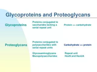
Glycoproteins • A glycoprotein is composed of a protein and a carbohydrate. The carbohydrate is attached to the protein in a postranslational modyfication. • The addition of sugar chains can happen either at asparagine, and is termed N-glycosylation, or at hydroxylysine, hydroxyproline, serine, or threonine, and is termed O-glycosylation. • Glycosylation is often present in proteins that are at least in part located in extracellular space. • The sugar group can assist in protein folding or improve its stability. • Glycoproteins are important for immune cell recognition.
Main pathways of enveloped virus entry pH-dependent pathway • Binding of viral glycoprotein with cell surface receptor the virus triggers receptor mediated endocytosis • In the cytoplasm the interior of the endocytic vesicle acidifies and the pH change triggers conformational change in the viral glycoprotein which leads to the fusion of the vesicle membrane with the viral envelope • The virus is released into the cytoplasm. Steven AC and Spear PG. Science VOL 313, 2006
Main pathways of enveloped virus entry pH-independent pathway • Upon binding of viral glycoprotein with cell surface receptor the viral glycoprotein undergoes conformational change leading to the fusion of viral envelope with the plasma membrane. Steven AC and Spear PG. Science VOL 313, 2006
Viral glycoprotein structure Viral glycoproteins are composed of three parts: • External domain – interacts with host • Transmembrane segment – spans the viral envelope membrane, typically -helix, • The endodomain – internal part Steven AC and Spear PG. Science VOL 313, 2006
Classes of ectodomains Class I • Found in othomyxoviruses, paramyxoviruses, retrovoruses, filoviruses and coronaviruses. • Form trimers with triple coiled-coil stem which in the postfusion state gives a distinctive six-helix bundle. • The active fusogenic form is obtained through a proteolytic cleavage which reveals the hydrophobic fusion peptide. Class II • Found in flaviviruses and alphaviruses • Initially are present in form of dimers but at low pH the dimers dissociate exposing the fusion loops. The fusion loops insert into host membrane which triggers trimerization of the glycoproteins. Other types of ectodomains that do not fit in class I and II. • Glycoprotein B of Herpes Simplex Virus (HSV) gB • Glycoprotein G of Vesicular Stomatitis Virus (VSV) Steven AC and Spear PG. Science VOL 313, 2006
Proposed mechanism for membrane fusion by class I fusion proteins. • After binding to a receptor on the cellular membrane, or on exposure to the low pH found in intracellular compartments (endosomes), the protein forms an extended conformation and the hydrophobic fusion peptide (red) inserts into the target membrane. • Several trimers are thought to be involved. • Protein refolding begins. The free energy thereby released causes the membranes to bend towards each other. • Formation of a restricted hemifusion stalk allows the lipids in the outer leaflets of the membranes to mix. • Forming the final, most stable form of the fusion protein, with the fusion peptide and transmembrane domain anti-parallel to each other but in the same membrane. Theodore S. Jardetzky and Robert A. Lamb Nature 427, 307-308 (22 January 2004)
Proposed mechanism for membrane fusion by class II fusion proteins. (A) Pre- and post-fusion conformations of class II fusion proteins. • At neutral pH, class II fusion proteins form flat head-to-tail homodimers that are oriented parallel to the viral membrane (upper panel), whereas exposure to the acidic pH triggers the dissociation of native homodimers, and the irreversible formation of homotrimers of the fusion proteins (lower panel). (B) Proposed model for membrane fusion induced by flavivirus class II fusion proteins. • (a) the envelope protein E binds to a receptor at the cell surface (upper membrane). • (b) after internalization of the virion in an endosomal compartment, acidic pH triggers conformational changes in the E protein leading to dissociation of the homodimer, insertion of the fusion loop into the target membrane and trimerization of the E protein. • (c) Domain III shifts and rotates, causing the C-terminal portion of E to fold back toward the fusion loop. This induces membrane bending and leads to hemifusion. • (d) A fusion pore is formed when the protein acquires its most stable form. CécileVoisset, Jean Dubuisson,Biology of the Cell 96 (2004) 413–420
HSV attachment and entry • HSV has a dsDNA genome which encodes for over 80 genes. • The envelope contains 12 different glycoproteins, five of which participate in virus entry gC, gB, gD, gH and gL, and only the last four take part in fusion of viral envelope with cell membrane. • The virus attaches to the cell through non-essential interaction of gC with heparan sulphate proteoglycan and through essential interaction of gD with with one of there receptors, either nectin-1, herpesvirus entry mediator (HVEM), or a specifically modified heparan sulphate. • Binding of gD to the receptor trigerrs a conformational change in gD leading to the release of C-terminal segment of the ectodomain from a strong intramolecular contact. • The free C-terminus of gD then interacts with gB or gH/gL complex to trigger conformational change leading ultimately to fusion. Heldwein EE. Science VOL 313, 2006 Spear PG. Cellular Microbiology 6(5), 2004.
Crystal structure of HSV glycoprotein B (gB) • HSV gB ia a 904 residue protein. • gB is composted of three protomers. • The residues Ala111-Thr669 coil around each other in a left handed coil. The protomers are further stabilized by multiple contacts that contribute do the trimer stability. • Moreover, each domain has 10 Cys residues that form 5 intramolecular disulfide bonds. • Each protomer is divided into five domains. Heldwein EE et al. Science VOL 313, 2006
Structure of HSV glycoprotein B Domain I • Contains a fold characteristic to pleckstrin-homology (PH) domain ( sandwich with -sheets of four and three strands) and another subdomain composed of four strand -sheet, short helix, hairpin and two-strand -sheet. Domain II • Contains six-strand barrel with a one helix. Domain III • Contains a long 44 residue helix, followed by short helix and four-strand -sheet. • Contributes to many essential trimer contacts. Domain IV • Contains two segments linked by disulfide bond. Domain V • The elongated domain fits into the groove between core domains of the two other protomers which reinforces the trimer interactions. Heldwein EE et al. Science VOL 313, 2006
Schematic model of gB conformation change • Transmembrane regions are shown as brown cylinders. • The linkers leading into and out of the domain I-II module would permit a large-amplitude rotation. • The structure described here might represent either a prefusion or a postfusion conformation. Heldwein EE et al. Science VOL 313, 2006
Crystal structure of VSV glycoprotein G • VSV has a ssRNA genome which encodes for only five genes. • Its envelope contains ~400 trimers of glycoprotein G. • G interacts with cellular receptors which leads to endocytosis. Then G is responsible for a pH induced fusion of the endocytic membrane with viral envelope. • G can adopt three conformational states. The first native state can be observed in pH 7. Then in the endocytic vesicle when the pH is lowered G changes conformation to the activated hydrophobic state which mediates the fusion. Then the third conformation is the post fusion form and is and inactive form. • G is composed of about 495 amino acids, it folds into 4 domains. The second domain is involved in trimerization. Roche S et al. Science VOL 313, 2006
Structure of VSV glycoprotein G Domain I • Made of three antiparallel -sheets • Glycosylation at position 320. • Antigenic site located in this domain. Domain II • Made of three segments with 4 -helices. • In the trimer the two longest helices form a six-helix bundle that resembles the structure found in class I fusion proteins. Domain III • Contains four -sheets and two helices. • Contains a disulfide bridge that stabilizes the domain. Domain IV • Has an extended -sheet structure that forms small six-stranded barrel at one end and three-stranded -sheet at the other end. • The barrel is glycosylated at position 163 and it’s stabilized by one disulfide bridge. • The two terminal loops constitute the membrane-interacting motif. • That domain is similar to the domain of class II fusion proteins. Roche S et al. Science VOL 313, 2006
Conformational change of the protein • pH induced conformational change is reversible Prefusion form postfusion form • In low pH the acidic amino acids in the six-helix bundle (domain II) are protonated and form hydrogen bonds leading to the transition to the postfusion form. • In higher pH the amino acids will be deprotonated leading to repulsive forces that destabilize the trimers and induce the transition to the prefusion form. The trimer • The core of the trimer is the six-helix bundle formed by domains II. • The stabilizing interactions are mostly hydrophobic with some salt bridges. Roche S et al. Science VOL 313, 2006