Download
1 / 33
2k likes | 6.97k Vues
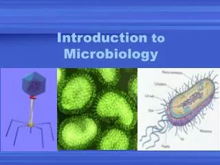
Introduction to Medical Microbiology. Dr Andrew Dodgson. Aims. What is medical microbiology? Why is it relevant? Some important concepts. Basic classification of organisms. Classifying bacteria. What is Medical Microbiology?.

E N D
Introduction to Medical Microbiology Dr Andrew Dodgson
Aims • What is medical microbiology? • Why is it relevant? • Some important concepts. • Basic classification of organisms. • Classifying bacteria.
What is Medical Microbiology? “the study of microorganisms (including bacteria, viruses, fungi and parasites) which are of medical importance and are capable of causing diseases in human beings”
What is Medical Microbiology? What organisms cause infection? How they cause infection. How to treat them. How to prevent infection.
Why is it Important? • Infection is one of the most important causes of mortality and morbidity in the population. • Approximately 30% of hospital patients are on antibiotics at any one time • 1 in 10 patients acquires an infection whilst in hospital.
A Few Concepts • Normal Flora • Contamination • Colonisation • Infection
Normal Flora • Human beings are not microbiologically sterile. • We are ALL covered with bacteria, fungi and some parasites. • Skin, nose, mouth, gastrointestinal tract … • ~109 bacteria per gram of faeces • Each person carries more non-human cells on their body than their own
Normal Flora Why? • Fulfil a range of useful functions (symbiotic in some cases). • Prevent other, pathogenic bacteria from gaining a foothold: • by taking up space • competing for nutrients. • In the gut they aid digestion & produce essential vitamins (folic acid & vitamin K). • Can cause disease IF they get into the wrong site- e.g. perforated appendix.
Contamination • Presence of an organism in a culture that was not in the sample when taken. • e.g. a culture of blood contaminated with an organism from the skin • sample contaminated in the lab
Colonisation • Presence of an organism at a site but not causing a tissue reaction (inflammation), symptoms or disease. • Could be normal flora • Could be abnormal flora-such as after the patient has received antibiotics.
Infection • where organisms invade a body site and their multiplication initiates a tissue reaction producing symptoms/disease.
Classification of Organisms • All living organisms are classified into: • Kingdom • Phyllum (family) • Genus • Species • Organisms that can cause disease are many and varied and include: • Viruses • Bacteria • Fungi • Parasites
Relevance of Classification • Different: • Diseases • Modes of transmission • Treatment-e.g. antibiotics don’t cure viral infections
Viruses Small (50-300nm) Unable to replicate independently Invade host cells and use their cellular machinery to replicate Influenza, Chickenpox (varicella), Herpes, Rhinovirus, HIV/AIDS Often difficult to treat
Bacteria • 500-800nm • Capable of independent replication • Cause of most infections seen in hospital • Pneumonia, bacterial meningitis, cellulitis, UTI… • Many different species • Treated with antibiotics
Fungi • Complex, large organisms • Eukaryotes (as are humans!) • Divided into yeasts & moulds • Cause a range of diseases e.g.: • Thrush • Athletes foot • Invasive & allergic aspergillosis • Many diseases are opportunistic.
Classifying Bacteria Why bother? Different bacteria: • cause different diseases • are susceptible/resistant to different antibiotics • some bacteria are common normal flora whilst other closely related species are pathogens
Classifying Bacteria How? • 1st into broad groups based on microscopic appearance • Then divided into species based on a range of different properties-often biochemical reactions e.g. some may be able to metabolise a sugar that others cannot.
Gram Stain Method of differentiating bacteria. Can be either Gram +ve or Gram –ve depending on how they appear with the stain. Can then be further grouped based on shape (rod=long thin or coccus=round). Thus we end up with 4 combinations: G+ rod, G+ coccus, G- rod, G- coccus
Gram Stain • STAIN the slide with crystal violet for 1-2 min. • Flood slide with Gram's iodine for 1-2 min. • Decolourise by washing the slide briefly with acetone (2-3 seconds). • Stain with safranin counterstain for 2 min. • View under microscope G+ve G-ve
Gram Stain Gives an initial idea of the possible identity of the organism. Can be done without growing the organism (i.e. rapid result) Thus can be done on pus, joint fluid, sputum, CSF 1st result available on blood cultures
Gram Stain Relevance of Gram reaction. • Gram +ve and gram –ve organisms ae susceptible to different groups of antibiotics. • Cause different diseases • Differ in their ability to survive in the environment-cleaning, infection control, outbreak management.
GPC • Clusters: usually characteristic of Staphylococcus spp., such as S. aureus • Chain or pairs: usually characteristic of Streptococcus spp., such as S. pneumoniae
GPR • Thick : usually characteristic of Clostridium spp., such as C. perfringens, C. difficile,C. tetani • Thin: e.g. Listeria spp.
GNC • Diplococci: usually characteristic of Neiseria spp., such as N. meningitidis or N. gonorrhoea. ThoughIn addition, Moraxella spp. and Acinetobacter spp.are often diplococcal in morphology. • Coccobacilli: usually characteristic of Acinetobacter spp., which can be either Gram-positive or Gram-negative, and is often called Gram-variable.
GNR • Thin rods: usually characteristic of enterobacteriaceae (coliforms), such as E. Coli • Coccobacilli: usually characteristic of Haemophilus spp., such as H. influenzae
GNR • Curved: usually characteristic of Vibrio spp.or Campylobacter spp., such as V. cholerae, C. jejuni • Thin needle shape: usually characteristic of Fusobacterium spp.
What can you see on the slide? • Gram +ve cocci • Gram +ve bacilli • Gram –ve cocci • Gram –ve bacilli Staphylococcus aureus – 100x
What can you see on the slide? • Gram +ve cocci • Gram +ve bacilli • Gram –ve cocci • Gram –ve bacilli Streptococcus pneumoniae
What can you see on the slide? • Gram +ve cocci • Gram +ve bacilli • Gram –ve cocci • Gram –ve bacilli Pseudomonas aeruginosa
Medical Microbiologists Medically qualified (mostly) Don’t spend time looking down microscopes… Clinical liaison- • interpretation of results • advice re Abx • appropriate testing MDT’s (ICU, Haematology, Renal, GUM…) Infection control