Cerebral Venous Thrombosis
200 likes | 936 Vues
Cerebral Venous Thrombosis. Department of Neurosciences Canberra Hospital March 1999. Cerebral Venous Thrombosis.

Cerebral Venous Thrombosis
E N D
Presentation Transcript
Cerebral Venous Thrombosis Department of Neurosciences Canberra Hospital March 1999
Cerebral Venous Thrombosis • Rare and severe disease characterised clinically by headache, papilledema, seizures, focal deficits, coma and death; pathologically by hemorrhagic infarction often contraindicating anticoagulation.
HISTORICAL BACKGROUND • Ribes 1825 • 45 yo man • 6 months severe headache, epilepsy and delirium. • Postmortem: superior sagittal sinus, left lateral and left parietal cortical vein thrombosis • Abercrombie 1828 • Postpartum cerebral venous thrombosis
INCIDENCE • Unknown incidence • Increased frequency of diagnosis since advent of DSA, CT & MRI/V. • Ehlers & Courville 1936 • 16 sagittal sinus thrombosis in 12500 autopsies • Kalbag & Woolf • 21.7 deaths per year in England & Wales from 1952 – 1961. • Male/female ratio = 1.29/1 • Males uniform age distribution • Females 61% CVT in 20-35 age group
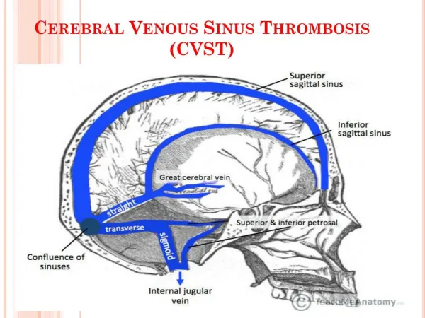
FREQUENCY OF VENOUS SITES(OFTEN MULTIPLE) • Superior sagittal sinus 72% • Lateral sinus 70% • Right 26% • Left 26% • Both 18% • Straight sinus 14.5% • Cavernous sinus 2.7% • Cerebral veins 38% • Superficial 27% • Deep 8% • Cerebellar veins 3%
FREQUENCY OF VENOUS SITES(SINGLE SITE) • One sinus only 23% • Superior sagittal sinus 13% • Lateral sinus 9% • Straight sinus 1% • Deep veins only 1% • Isolated cortical veins 1%
ETIOLOGY • IDIOPATHIC • INFECTIVE • Local: direct septic trauma • Intracranial infection • Regional infection • General: Septicemia, measles, encephalitis, HIV, CMV, malaria • NONINFECTIVE • Local: head injury, neurosurgery, tumors, infusions into jugular vv • General: Postoperative, pregnancy/postpartum, dehydration, inflammatory bowel disease, connective tissue disease, malignancy, thrombophilia.
CLINICALLY BY SYNDROMIC DESCRIPTION • 1.Isolated intracranial hypertension 40% • mimic benign intracranial hypertension • 2.Focal signs 50% • 3.Cavernous sinus thrombosis • 4.Unusual presentations • Psychiatric disturbances, migraines, subarachnoid hemorrhages.
CLINICALLY BY SYMPTOMATOLOGY • Headache 75% • Papilledema 49% • Motor or sensory deficit 34% • Seizures 37% • Drowsiness, mental changes, confusion, or coma 30% • Dysphasia 12% • Multiple cranial nerve palsies 12% • Cerebellar incoordiantion 3% • Nystagmus 2% • Hearing loss 2% • Bilateral or alternating cortical signs 3%
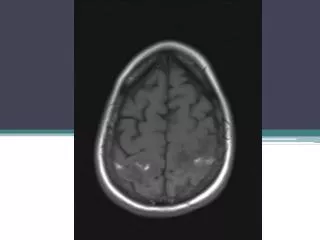
INVESTIGATIONS – DIAGNOSTIC • .CT • Infarction in nonarterial distribution (often hemorrhagic) • Empty delta sign • Dense triangle sign • Cord sign • .DSA • .MRI/V • Early: absence of flow void & isointense on T1 for occluded vessel; Hypointense on T2 • Late:hyperuintense thrombus on T1 & T2 • .CRANIOTOMY • OTHERS: EEG, CSF, isotope brain scanning.
INVESTIGATIONS – ETIOLOGIC • FBE • ANA, antiphospholipid antibodies • APC resistance (Factor V Leiden) • Antithrombin • Protein C, S • Homocysteine • Prothrombin gene mutation • Repeat tests in 4-6 months.
TREATMENT • 1.Infective cause • 2.Increased intracranial pressure • 3.Anticoagulation: • initially heparin • warfarin (?duration) • direct urokinase infusion
PROGNOSIS • MORTALITY • Untreated: 50% • Treated: nonseptic cause 10% • septic cause 30% • OUTCOME • 77% no sequelae • 20% develop thrombosis intra or extracerebrally • Longest followup study is 8 yrs.
SUMMARY • Uncommon but life threatening disease. • Mimic many benign conditions. • Untreated carries 50% mortality. • If treated, majority of patients have no long term disability. • An underlying cause should always be sought.
THROMBOPHILIA & CEREBRAL VENOUS THROMBOSIS • 25% CVT have a detectable thrombophilia (APC resistance; antithrombin, protein C or S deficeincy, antiphospholipid syn) • 20% CVT have APC resistance • 95% APC resistance due to Factor V leiden. • In patients with CVT attributable to APC resistance, • 72% had a second contributing factor (OCP, other thrombophilia) • Contribution of G20210A prothrombin gene mutation unknown.
ORAL CONTRACEPTIVE PILL AND CVT • Relative risk of developing CVT • OCP RR13 • Thrombophilia RR4 • OCP & Thrombophilia RR30 • De Bruijn et al. Case control study of risk of cerrebral sinus thrombosis in oral contraceptive users who are carriers of hereditary prothrombotic conditions. BMJ 1998: 316, 589-92.
ISSUES • APC resistance is not always caused by Factor V Leiden. • Different thrombogenicity of second vs third generation OCP. • Contribution of G20210A prothrombin gene mutation to CVT.