Download
1 / 23
230 likes | 436 Vues
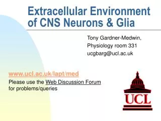
Extracellular Environment of CNS Neurons & Glia. Tony Gardner-Medwin, Physiology room 331 ucgbarg@ucl.ac.uk. www.ucl.ac.uk/lapt/med Please use the Web Discussion Forum for problems/queries. CNS Extracellular Environment Is there any e-c space? - size, composition

E N D
Extracellular Environment of CNS Neurons & Glia Tony Gardner-Medwin, Physiology room 331 ucgbarg@ucl.ac.uk www.ucl.ac.uk/lapt/med Please use the Web Discussion Forum for problems/queries
CNS Extracellular Environment Is there any e-c space? - size, composition The macro-environment: CSF, blood Homeostasis, Disturbances - (normal, pathological) Role of glia in K+ homeostasis Failures of regulation: The war between + feedback and - feedback From Neuron to Brain (Nicholls, Martin & Wallace) - Chapter on Neuroglia
Is there any extracellular space? Photoreceptors (dark) and glial cells in the compound bee eye
Measurement of Extracellular Space Volume 1. 15-20nm gaps => 2% - 5% but ? are cells swollen to occlude space? 2. Try e-c markers from blood: don’t show up ( ‘Blood-Brain Barrier’ ) ? Is this lack of penetration due to zero e-c space? 3. Markers from CSF: 10-20% space, but v slow equilibration 4. Improved EM technique: rapid freezing -> 18 - 25% Measurement of Extracellular Space Volume 1. 15-20nm gaps => 2% - 5% but ? are cells swollen to occlude space? 2. Try e-c markers from blood: don’t show up ( ‘Blood-Brain Barrier’ ) ? Is this lack of penetration due to zero e-c space? 3. Markers from CSF: 10-20% space, but v slow equilibration 4. Improved EM technique: rapid freezing -> 18 - 25%
EM after rapid freezing, <30s after cessation of circulation
EM after rapid freezing, 8 min after cessation of circulation
Measurement of Extracellular Space Volume 1. 15-20nm gaps => 2% - 5% but ? are cells swollen to occlude space? 2. Try e-c markers from blood: don’t show up: ‘blood-brain barrier’ ? Is the lack of penetration due to zero e-c space? 3. Markers from CSF: 10-20% space, but v slow equilibration 4. Improved EM technique: rapid freezing -> 18 - 25% 5. Release of e-c markers from electrodes, and measurement of concentration, with ion-selective electrodes -> 15 - 25%
Measurement of Extracellular Space Volume Five methods …… Conclusions ~ 20% e-c space (similar to rest of body) Cut off from blood (unlike elsewhere) Free (but slow) diffusion exchange with CSF ….. but what about its composition?
Functions: Mechanical: floating brain ↓ postural effects lubrication / movement pressure ≈venous (10 mmHg) Variable volume reservoir (but only a few % of brain volume) Clearance (like lymph) Homeostasis: regulated composition
How successful is K+ homeostasis? Plasma [K+] CSF [K+] Normal Diet: 4.2 2.8 mM Low K diet: 1.6 2.7 mM High K diet: 7.1 3.0 mM Baseline e-c [K+]o measured with ion-selective micro-electrodes is similar to CSF (normally less than plasma, and well regulated)
Does neural activity alter e-c [K+] ? Epileptic discharges in cortex -> transient increase of extracellular [K+] and depolarisation of astrocytes
Electrical properties of glial cells Initially studied in leech n.s. 1. Large resting potentials ( ~ -90 mV cf neurons -70 mV) 2. Inexcitable (no action potentials) 3. Electrically coupled (via GAP junctions) 4. Vm sensitive to [K+]o - follows Veq(K+) - membranes very selectively permeable to K+ 5. Slow, long depolarisations when adjacent neurons are stimulated 6. Can take up K+, GABA, glutamate from e-c space
Effect of light stimulation on K+-selective electrode concentration measurements in bee eye [K+]i (mM) [K+]i [K+]o
Effect of visual stimulation with moving bars of light, on glial Vm in cat visual cortex -60 -72 Glial membrane potential (mV) 0 10 20 30 40 50 s
K+ ‘spatial buffer’ mechanism disperses potassium from regions of activity & build-up, into normal tissue and to surface fluid Diagrammatic version of the coupled astrocyte network
REGULATION OF E-C [K+] 1. EXCHANGE WITH BLOOD: SLOW !! Accurate homeostasis but only regulation of average concentration over many hours 2. DIFFUSION : Evens out local disturbances, reducing their maximum effect. Effective only over short distances, and would be BETTER without the blood-brain barrier. 3. GLIAL UPTAKE and DISPERSAL via SPATIAL BUFFER mechanism. Assists dispersal by diffusion ( ~ 5x) Helps to reduce disturbances due to neural activity. But why is regulation important ??
Wave of drastic, but transient disturbance of extra-cellular K+ and Ca++ concentrations spreading from local trauma in baboon cortex ‘Cortical Spreading Depression’ (probably part of the syndrome in migraine and stroke)
- feedback + feedback Positive feedback may lead to e-c instability in stroke, migraine and trauma Raised [K+]o Depolarisation GLIA NEURONS Spatial Buffer currents K+ uptake and dispersal Transmitter Release Raised PNa, PK, PCl K+ release