Stroke
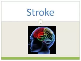
Stroke. Carla Kreft ND LAc MSOM. What is a Stroke?. Short version: Circulation to the brain is cut off, the neurons are deprived of oxygen, the neurons die The person is left with symptoms that correspond to the area of the brain that died. First Signs of Stroke.

Stroke
E N D
Presentation Transcript
Stroke Carla Kreft ND LAc MSOM
What is a Stroke? • Short version: • Circulation to the brain is cut off, • the neurons are deprived of oxygen, • the neurons die • The person is left with symptoms that correspond to the area of the brain that died.
First Signs of Stroke • symptoms such as weakness, speech disturbance, numbness and loss of vision. Without the prompt restoration of blood flow, permanent brain damage occurs
Abnormal sense of taste Change in alertness (level of consciousness) Apathetic, withdrawn Sleepy, lethargic, stuporous Unconscious, comatose Difficulty speaking or understanding speech Difficulty swallowing Difficulty writing or reading Headache May occur when lying flat May awaken patient from sleep May increase with change in position May increase with bending, straining, and coughing Loss of coordination Loss of balance Movement changes Difficulty moving any body part Hand tremor Loss of fine motor skills Weakness of any body part Nausea, vomiting Seizure Sensation changes Abnormal sensations Decreased sensation Facial paralysis Numbness or tingling Vision changes Any change in vision Decreased vision, loss of all or part of vision Double vision Eyelid drooping Pupils different size Uncontrollable eye movements Symptoms of a Stroke
Blood Circulation from the Heart TO the Brain • Aorta > Brachiocephalic, Common Carotid, Subclavian > Internal Carotid & Vertebral Artery
Arteries of the Brain Notice the: • Anterior Cerebral Artery • Middle Cerebral Artery • Posterior Cerebral Artery
2 Types of Stroke • Ischemic - 83% • Hemorrhagic - 17%
Stroke Mortality • 30 day mortality rates among people 45 - 64 yo • Ischemic stroke: 8%- 12% • Hemorrhagic strokes 37% - 38% • Stroke is the 4th leading cause of death in the USA
Ischemic Stroke (80%) • In an ischemic stroke, a blockage occurs in a blood vessel, depriving the area distal to the blockage of oxygen. • Thrombotic Stroke- a clot,or, thrombus, forms within a blood vessel in the brain until it gets so big that it blocks blood flow. • Embolic Stroke - an object travels in the bloodstream until it gets stuck and blocks blood flow. • Lacunar or Small vessel stroke
Thrombotic Strokes • Thrombi are blood clots. They frequently form within an unhealthy, atherosclerotic vessel, on top of an atheroma. They can impede blood flow if they get too large (atherothrombotic stroke). • Atheromas, or, fatty plaques, can occur in any major cerebral artery and are common at areas of turbulent flow, particularly at the carotid bifurcation. If the cap on the atheroma ruptures or ulcerates, it causes thrombus formation.
Atherosclerosis • excess LDL passes through endothelial cells to tunica media. • Becomes oxidized & toxic. • Macrophages try to remove it & become foam cells • Dead macrophage lipids create a lipid core • A cap of collagen & elastin covers the plaque • If it is unstable, the plaque ruptures due to turbulent flow of blood & a thrombus forms
Vascular inflammation from disorders such as: acute or chronic meningitis vasculitic disorders syphilis Dissection of intracranial arteries or the aorta (leads to clot formation at site) Hypercoagulability disorders eg Antiphospholipid syndrome Hyperhomocysteinemia Hyperviscosity disorders eg: polycythemia thrombocytosis hemoglobinopathies plasma cell disorders Rare disorders eg: moyamoya disease Binswanger's disease Older oral contraceptive formulations increase risk of thrombosis FYI:Thrombi tend to occur during the night and thus are first noticed on awakening Less Common Causes of Thrombosis (clotting) Include:
Small Vessel or Lacunar Strokes • Small vessel-related strokes, are also called “lacunar” infarctions due to their lake-like appearance on brain imaging • Usually caused by thrombosis of small penetrating arteries. due to Atherosclerosis • Small arteries are particularly susceptible to injury from smoking, hypertension, and diabetes. They eventually thrombose with platelet-rich clots.
Lacunar Infarcts • Although area of brain injured is much smaller than in most large vessel strokes, many key motor and sensory pathways run through the deep, small-vessel territory, often resulting in significant symptoms despite the smaller size of injury
Embolic Stroke Emboli commonly originate from: • Ulcerated plaques at the carotid bifurcation in the neck or the aortic arch • Cardiac thrombi, (cardioembolic stroke) especially in the following conditions: • Atrial fibrillation (15-20% of ischemic strokes yearly) • Rheumatic heart disease (usually mitral stenosis) • Post-Myocardial Infarction • Vegetations on heart valves from bacterial or non-bacterial endocarditis • Prosthetic heart valves
Clots that form and dislodge after open-heart surgery or other invasive cardiovascular procedures eg, catheterization Rarely, emboli consist of: Fat (from fractured long bones), Air (in decompression sickness) Venous clots that pass from the right to the left side of the heart through a patent foramen ovale with shunt (paradoxical emboli). Rarely, thrombosis of the subclavian artery results in embolic stroke in the vertebral artery or its branches. Other sources of Emboli
Core Ischemic Zone & Ischemic Penumbra • Core Ischemic Zone: area of irreversible ischemia and neuronal cell death • Blood flow falls below 10% - 25% of normal • Ischemic Penumbra: area of reversible ischemia • Blood flow falls to 25%-50% of normal. • Normal CBF:50-60 cc/100 g/minute (14% of CO) • Less electrical activity:CBF 20-30cc/100g/min • Penumbra: 10-18cc/100g/min • Neuronal metabolism stops (death): CBF <10 cc/100g/min
Ischemic Penumbra • Ischemic Penumbra: The ischemic zone that surrounds a central core of infarction. • At <18cc/100g/min there is a loss of electrical activity in the neuron. • Here,the right Middle Cerebral Artery is occluded. Collateral circulation from the right Anterior Cerebral Artery distributes variable amounts of blood to the right MCA territory. • Penumbra may be clinically symptomatic but can be reversed if blood flow is restored within 3-6 hours
Window of Opportunity • Viability of brain tissue is preserved if perfusion is restored within a critical time period (2 to 4 hours)
The Neuron • Resting Membrane Potential (Na+ outside, K+ inside. -55mV) • Stimulus - causes Na+ channels to open • Threshold - cell membrane depolarizes to +35mV • Action Potential (+20-50mV) Propagation (Na+ rushes into neuron along axon) • Neurotransmitter release at axon terminals into synapse • Repolarization - Na channels close, K+ channels open, K+ rushes out of cell • Na+/ K+ pump restores Na to outside of cell & K+ to inside of cell
Aerobic & Anaerobic Respiration • In the presence of oxygen, a cell (neuron) makes 36 ATP from 1 glucose via citric acid cycle & oxidative phosphorylation • In the absence of oxygen, the cell can only make 2 ATP via glycolysis, or fermentation
Cellular Effects of Oxygen Deprivation • During an ischemic stroke, neurons are deprived of oxygen, carried by the red blood cells. • Without oxygen, the hypoxic cell can only generate ATP through anaerobic respiration, or glycolysis. • A by-product of glycolysis is lactate, or lactic acid, which increases the acidity of the blood. • The neurons cannot make enough ATP to power the Na/Kpump and keep the Na outside of the cell.
Cytotoxic Edema • Cytotoxic edema is caused by entry of sodium (Na) and other solutes into the neuron • Cytotoxic edema within endothelial cells, allows intravascular Na to traverse the capillary wall and replenish sodium in the extracellular space. This is called Ionic edema. • After 3-6 hours of ischemia, vasogenic edema results from breakdown of tight junctions between endothelial cells, allowing extravasation of serum proteins and water into the brain • The end result is neuronal death, loss of BBB and brain swelling
Cytotoxic Edema • Cytotoxic edema is reversible at the stage where the Na/K pump is dysfunctional from lack of O2 & ATP • When organelles like mitochondria become damaged, it the degeneration can no longer be reversed. • The area becomes edematous and ultimately fibrosed
Gliosis (Fibrosis) • Astrocyte extensions create a dense web to fill in the empty space generated by dead neurons. • Microglia clean up the debris & secrete may cytokines to stimulate endothelial cells to create new vessels and fibroblasts to lay down collagen. • Scar tissue restores the chemical integrity of the blood-brain barrier.
Timeline: Histopathological Changes during different phases of stroke
Recap of Cellular Effects of Ischemic Stroke • A blood vessel in the brain becomes blocked, due to a thrombus or an embolus • Blood / oxygen no longer reach the neurons • Neurons switch over to anaerobic glycolysis, making much less ATP and lactate • Lactic acid accumulates and intracellular pH decreases • Without enough ATP, the Na/K ion pump fails. • Sodium, and thus water, and calcium enter the cell & it swells (cytotoxic edema). This trapped fluid can be visualized on diffusion weighted MRI. • If oxygen is not restored to the area, mitochondrial function ceases, which signals neuronal death and membrane lysis occurs • Gliosis creates a fibrotic scar, visible as an area of increased signal density on MRI
Hemorrhagic Stroke (20%) • In hemorrhagic stroke, a blood vessel in the brain breaks or ruptures. • Occurs in 2 places: • A blood vessel inside the brain tissue: intracerebral hemorrhage (ICH) • A blood vessel on the surface of the brain that bleeds into the space between the brain and the skull: subarachnoid hemorrhage (SAH)
Intracerebral Hemorrhage (ICH) ICH is most commonly caused by: • HYPERTENSION (60-70% of ICH) • Arteriovenous malformations & aneurysms • Head trauma • Tumors • Infection • Sympathomimetic drugs eg cocaine, amphetamines
Hypertension & ICH • ICH is more likely to result in death or major disability than ischemic stroke or subarachnoid hemorrhage. (800,000 people /year. 5.4 million stroke survivors. $73 billion in costs) • Chronic hypertension produces a small vessel vasculopathy characterized by lipohyalinosis, fibrinoid necrosis, and development of Charcot-Bouchard aneurysms, affecting penetrating arteries throughout the brain.
Common locations for Hypertensive ICH • Hypertensive-related ICH can occur in any location in the brain but has a particular predilection for: • the basal ganglia (40-50%), especially the putamen (green) • the thalamus (blue) (10-15%) • the cerebellum (5-10%) • the pons in the brainstem (5-12%)
Arteriovenous Malformation (AVM) • Arteriovenous malformations (AVMs) are abnormal tangles of blood vessels that can be asymptomatic prior to rupture and are usually diagnosed via angiography
Amyloid Angiopathy - deposition of an abnormal protein weakens blood vessel walls. usually a disease of the elderly Coagulopathy - increased bleeding tendency. from disorders such as liver disease, malignancy, or blood thinning medications Ischemic stroke with secondary hemorrhage Vasculitis -Inflammatory disorders involving the blood vessels of the Less common causes of ICH
Subarachnoid Hemorrhage (SAH) • Subarachnoid hemorrhage results from the bleeding of an artery, usually around the base of the brain. • Least common type of stroke: ~ 5% of all strokes. • HEAD TRAUMA is the most common cause of SAH. Falls in the elderly & motor vehicle accidents in the young.
Traumatic SAH • Traumatic brain injury results from an external force hitting the head at high velocity which causes the brain to hit the inside of the skull, damaging blood vessels in the subarachnoid space. • This can be life threatening if the intracranial pressure exceeds the mean arterial blood pressure
Aneurysm • 80% of non-traumatic subarachnoid hemorrhage results from a ruptured berry ANEURYSM. • Aneurysms are defects in blood vessels thought to expand as a result of hydrostatic pressure from pulsatile blood flow and blood turbulence, which is greatest at the arterial bifurcations
Trauma is the most common. Usually falls in the elderly & motor vehicle accidents in the young Ruptured Berry Aneurysm 80% of non traumatic SAH Other causes: Bleeding from an arteriovenous malformation (AVM) Bleeding from a cerebral aneurysm Bleeding disorder Use of blood thinners Other Causes of SAH
Risk Factors for SAH high blood pressure cigarette smoking oral contraceptives pregnancy and child birth cocaine abuse aneurysm fibromuscular dysplasia (FMD) and other connective tissue disorders history of polycystic kidney disease (from htn) Symptoms of SAH Main symptom is a severe headache that starts suddenly and is often worse near the back of the head. Patients often describe it as the "worst headache ever” Blood throughout the subarachnoid space,causes headache and neck stiffness Raised intracranial pressure, causes possibility of depressed conscious level, headache, vomiting, papilloedema Risks & Symptoms for SAH
Subarachnoid Anatomy • The subarachnoid space lies just exterior to the the brain tissue & its covering (the pia mater) • Normally, this space is full of CSF to cushion and protect the brain.
Diagnosis: Neurological exam - Is it a stroke? • Stroke can be clinically diagnosis based on history and physical examination (NIH Stroke Scale evaluation). • http://www.strokecenter.org/professionals/stroke-diagnosis/stroke-assessment-scales-overview/ for complete chart evaluation tools LOOKING FOR SIGNS OF THESE AND MORE: • Aphasia: total or partial loss of ability to understand or use words - trouble finding words or unable to speak. problems understanding what others are saying or trouble with reading, writing or math. may have trouble talking yet understand what others say. • Apraxia: inability to control muscles making uncoordinated and jerky movements • Dysarthria: loss of control of muscles in face & mouth - voice may sound slurred, muffled, hoarse. mouth may droop on one side of face from muscle weakness. • Dysphagia: difficulty swallowing • Paralysis: loss of muscle function and sensation • Hemiparesis: weakness of muscles on one side of the body. • Hemianopia: loss of sight in half of visual field
Stroke Syndromes • The “hallmark” of an acute stroke is the sudden onset of focal neurologic dysfunction, corresponding to a distinct vascular territory. • A careful history and neurologic examination can often localize the region of brain dysfunction
Brain Lateralization • The brain is divided into 2 hemispheres: Left & Right. The 2 hemispheres are not the same. • Although they look similar to each other, in most people, only one side (usually the left) contains areas that allow a person to produce speech (Broca’s area) & to comprehend speech (Wernicke’s area). • E.g. In 95% of right-handers, the left side of the brain is dominant for language. Even in 60-70% of left-handers, the left side of brain is used for language. • Thus, a stroke on the left side of the brain will produce symptoms that are different from a stroke on the right side of the brain.
LEFT BRAIN FUNCTIONS uses logic detail oriented facts rule words and language present and past math and science can comprehend knowing acknowledges order/pattern perception knows object name reality based forms strategies practical safe RIGHT BRAIN FUNCTIONS uses feeling "big picture" oriented imagination rules symbols and images present and future philosophy & religion can "get it" (i.e. meaning) believes appreciates spatial perception knows object function fantasy based presents possibilities impetuous risk taking Left vs Right Brain Functions