Download
1 / 31
360 likes | 1.43k Vues
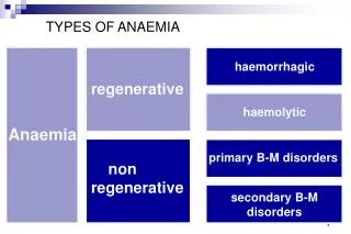
LABORATORY INVESTIGATION OF ANAEMIA. The full blood count (FBC) usually provides the first evidence of anaemia, e.g. low Hb. The peripheral blood film (PBF) may reveal red cell abnormalities characteristic of particular disorders, e.g. sickle cells. .

E N D
LABORATORY INVESTIGATION OF ANAEMIA The full blood count (FBC) usually provides the first evidence of anaemia, e.g. low Hb. The peripheral blood film (PBF) may reveal red cell abnormalities characteristic of particular disorders, e.g. sickle cells.
The reticulocyte count (retics) reflects the bone marrow's response to anaemia. A low retic count indicates bone marrow hypoplasia. Reticulocytosis (high retic count) indicates the marrow is still responding.
A bone marrow (BM) aspirate should be taken if a primary marrow disorder is suspected, e.g. myelodysplasia or leukaemia. Trephine is appropriate with infiltrative conditions, e.g. lymphoma or carcinoma.
Haematinic assays for serum ferritin, B12 and folate should be evaluated in all cases. Iron deficiency may mask the megaloblastic changes of B12 and/or folate deficiency.
IRON DEFICIENCY ANAEMIA (IDA) IDA is the commonest cause of anaemia throughout the world. Iron is an essential constituent of haemoglobin and hence RBCs require large amounts (compared to other cells). Symptoms are due to a deficit of Hb; the degree of anaemia depends upon the iron deficit.
Every cell in the body contains iron and loss of any cell from the body (skin, gut, etc.) results in iron loss, i.e. obligatory iron loss. There is also occult (mysterious) and sometimes overt (unconcealed) loss of red cells from body surfaces. Extra requirements to meet specific needs include growth, pregnancy or blood donation.
IRON METABOLISM Iron is present in the diet in many forms. Haem is the most important source. Vegans may need to supplement their dietary intake with non-organic iron. A normal adult requires 15-20 mg of iron per day to remain in balance.
Iron is normally absorbed by active transport across the wall of the duodenum and upper part of thejejunum. If large amounts of iron are ingested the active transport mechanism is overtaken by passive diffusion.. Disease of the upper small gut can lead to malabsorption of iron, e.g. coeliac disease or tropical sprue.
Iron is best absorbed in the ferrous (reduced) form (Fe++). Absorption is improved by reducing substances, e.g. ascorbic acid (vitamin C). Absorption is also increased by certain iron chelators and by alcohol.
Iron absorption is normally relative to the needs of the body. About 10% of dietary iron is usually taken up by the body but this can increase several-fold in iron deficiency, or reduce if the body has a surplus.
Most iron in the body is in the form of haem; present in large amounts in red cells, muscle and liver where it is essential for oxygen supply. Iron is also present in many enzyme systems, e.g. electron transport systems. The transport and storage of iron mainly involves three proteins: transferrin transferrin receptor (TfR) ferritin
Transferrin actively binds and transports iron in the body and can be estimated by measuring the serum total iron binding capacity (TIBC). Transferrin increases in iron deficiency and decreases with iron overload, liver disease, infection, malignancy and protein deficiency.
Hereditary Haemochromatosis • Autosomal recessive condition resulting in a congenital error in iron metabolism with increased iron deposition in tissues resulting in brown (bronzed) pigmentation of the skin and cirrhosis of the liver. Deposits in the pancreas lead to diabetes (bronzed diabetes). The gene involved is called HFE. • Haemosiderosis (iron overload) • Deposits of haemosiderin in tissues resulting from: • increased iron absorption • increased iron intake • transfusion siderosis
Excess iron is stored mainly in macrophages as haemosiderin; an insoluble protein-iron complex formed by lysosomal degeneration of ferritin. Haemosiderin is demonstrated by the Perl's (Prussian blue) staining reaction. Ferritin is the water soluble protein-iron complex formed when iron combines with apoferritin. Iron in ferritin is in the ferric form (Fe+++) and must be reduced before it can be utilised.
LABORATORY INVESTIGATION OF IRON DEFICIENCY ANAEMIA • Full Blood Count • Serum Ferritin • Serum Iron & Total Iron Binding Capacity • Serum Transferrin • Bone Marrow
FULL BLOOD COUNT can be suggestive but not diagnostic of iron deficiency. Negative iron balance produces microcytosis (low MCV) and hypochromasia (low MCH). Red cell morphology varies from mild anisocytosis to marked anisopoikilocytosis. Thrombocytosis is common. Leucocytes are usually normal.
SERUM FERRITIN is now a standard diagnostic test for IDA; only iron deficiency will give a low result. Normally the level of serum ferritin reflects the body iron stores (100 μg/L = 800 mg of iron). A value <15 μg/L is diagnostic of IDA.
Circumstances in which the serum ferritin is normal or high in the presence of IDA: • Liver dysfunction; ferritin is released when hepatocytes are damaged • Increased haem turnover; haemolysis and trauma (including surgery) • Inflammatory lesions; malignancy, infection and inflammation
Serum ferritin is usually assayed using enzyme-immunological procedures, e.g. EIA or ELISA.
SERUM IRON (SI) and TOTAL IRON BINDING CAPACITY (TIBC) In iron deficiency the SI is low (<10 μmol/L) and the TIBC is usually raised (>70 μmol/L). Erythropoiesis is iron-deficient when the transferrin saturation (SI TIBC x 100%) falls below 15%.
The SI shows marked diurnal variation. It may also be low in the presence of infection or inflammation. The TIBC is also affected by nutrition and may be low in malnourished persons despite iron deficiency. For these reasons the serum ferritin is now preferred. SI and TIBC are useful when the ferritin is falsely high, e.g. liver damage.
The serum iron can be quantitated based on the development of a coloured complex when ferrous iron is treated with a chromagen solution, e.g. ferrozine. The iron-binding capacity is usually measured by adding an excess of iron and measuring the iron retained after the action of a suitable binding reagent, e.g. light magnesium carbonate.
In the plasma, iron is bound to transferrin and the TIBC depends on the concentration of this protein. The transferrin to which iron is not bound is known as the unsaturated iron-binding capacity (UIBC). SI + UIBC = TIBC
REVIEW: Excess iron as ferric chloride is added to serum. Iron that does not bind to transferrin is removed with magnesium carbonate. The iron concentration of the iron-saturated serum is then measured.
SERUM TRANSFERRIN is measured directly by immunological methods (e.g. immunodiffusion, nephelometry,) rather than by its ability to bind iron. Normal serum contains 2-4 g/L of transferrin.
BONE MARROW specimens should be stained for iron (Perl's Prussian blue) as a routine. The main use of a marrow aspiration is in clarifying a differential diagnosis.
Stainable iron normally excludes iron deficiency. • Plentiful macrophage iron, but no sideroblasts, indicates a secondary anaemia. • Microcytosis and many sideroblasts indicates a haemoglobinopathy. • Ringed sideroblasts indicate sideroblastic anaemia.