Leukemia
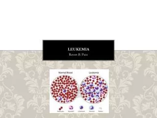
Leukemia. Leukemia is a general term for a group of malignancies of either lymphoid or hematopoietic cell origin. General features: 1- The number of circulating leukocytes is often greatly increased. In few cases the number is of normal range (aleukemic leukemia).

Leukemia
E N D
Presentation Transcript
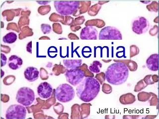
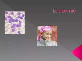
Leukemia Leukemia is a general term for a group of malignancies of either lymphoid or hematopoietic cell origin.
General features: 1- The number of circulating leukocytes is often greatly increased. In few cases the number is of normal range (aleukemic leukemia). 2- The bone marrow is diffusely infiltrated with leukemic cells, often with encroachment on normal hematopoietic cell development. 3- Consequent failure of normal leukocyte, red cell, and platelet production which results in anemia, infection, and hemorrhage. 3- Infiltration of leukemic cells in the liver, spleen, lymph nodes, and other organs is common.
Pathological features of leukemia 1- Bone marrow: -Shows leukoblastic reaction mainly in the sternum, ribs, vertebrae, and pelvis. - The leukemic tissue appears reddish brown in acute leukemia and grayish-white in chronic leukemia. 2- Blood changes: A- Increase of the total leucocytic count due to blood invasion by the leukemic cells. B- In acute leukemia the blast type of cells predominate, while in chronic leukemia more differentiated cells appear ( myelocytes, and lymphocytes).
C- Anemia especially in chronic leukemia. D- Thromocytopenia. 3- Leukemic infiltrate: - In the liver, adrenal, kidney…………and skin. The infiltrate begins around the blood vessels and as it increases in size it destroys the surrounding tissue. 4- Lymph nodes: - The infiltrate causes enlargement of the nodes and loss of the normal pattern. Affection of the perinodal tissue fuses the nodes together. 5- Splenomegaly: - Due to leukemic infiltrate and splenic infarcts caused by thrombosis of its vessels.
Etiologic factors The difficulty of pinpointing one cause of leukemias indicates that it may be caused by a combination of different factors: 1- Chromosomal translocations and oncogenesis: non-random karyotypic abnormalities, most commonly translocations are present in the majority of leukemias. 2- Inhereted genetic factors: Down syndrome (trisomy 21) and neurofibromatosis are associated with increased incidence of childhood leukemia.
3- Viruses: Three viruses HTLV-1, EBV, and HHV-8 have been implicated as causative agent of lympho-hemetopoietic neoplasms HTLV-1→ adult T-cell leukemia/lymphoma. EBV→ African Burkitt’s lymphoma. HHV-8 → Primary effusion lymphoma. 4- Iatrogenic factors: Radiotherapy and certain forms of chemotherapy used to treat cancer increase the risk of subsequent leukemia especially AML.
Classification: - The classification of leukemia is based on: 1- The predominent cell type: a- Myeloid leukemia. b- Lymphatic leukemia. c- Plasma cell and histiocytic leukemia (rare). d- Monocytic leukemia. 2- The degree of cellular differentiation: a- Acute leukemias (undifferentiated with rapid course). b- Chronic leukemias ( Moderately differentiated with slow course.
Acute leukemias General considerations: 1- A predominance of blasts and closely related cells in the bone marrow and peripheral blood is characteristic. 2- The most common malignancies of the pediatric age group, acute leukemias occur most often in children. They exhibit a second incidence peak in the late adult age. 3- Cytogenetic abnormalities are frequent. For example, the 9;22 translocation results in a morphologically unique chromosome, the Philadelphia chromosome (Ph 1). This abnormality, better known for its association with chronic myelogenous leukemia, is associated with a poorer prognosis when it occurs in acute leukemias. 4- Without therapeutic intervention, acute leukemia follows a short and precipitous course, marked anemia, infection, and hemorrhage, and death occurs within 6 to 12 months.
ACUTE LYMPHOBLASTIC LEUKEMIA (ALL) EPIDEMIOLOGY: Childhood. Most common childhood malignancy. PATHOGENESIS: The c-myc gene is involved, as in Burkitt Lymphoma. IMMUNOTYPES: Several different cell-types can yield ALL. Flow Cytometry can be used to identify the specific type. B-cell ALL: More common, better prognosis. Subtypes are Pre-Pre-B-ALL, Pre-BALL, and Mature B-ALL (worst prognosis) T-Cell ALL: More common in adolescents, worst prognosis. Null Cell ALL
SUBTYPES: 3 Subtypes proposed by the French-American-British (FAB) Group: • L1 LYMPHOBLASTS: Small, plain cells. CLINICAL: Best prognosis, common in the children 3-7 age group. • L2 LYMPHOBLASTS: Contain prominent nucleoli. CLINICAL: Found in infants younger than 1, or common in adolescents (T-cell immunotype) or in adults. • L3 LYMPHOBLASTS: Burkitt's Leukemia. Identical histology to Burkitt's Lymphoma. Larger cells with vacuoles in cytoplasm, and showing characteristic starry sky appearance . CLINICAL: Poor prognosis, found in children 6-11 years of age.
Clinically: Hepatosplenomegaly Generalized Lymphadenopathy, particularly cervical nodes. Normocytic normochromic anemia, thrombocytopenia, neutropenia. May have CNS involvement. PATHOLOGY: Numerous cases have cytogenetic abnormalities. Hyper-diploidy is a common abnormality and is a favorable prognostic indicator.
DIAGNOSTIC CRITERIA: - Numerous blast cells must be present in the bone marrow or peripheral blood. - TDT -Positive: Terminal Deoxynucleotidyl Transferase is present in the lymphoid cells, distinguishing it from the myeloid cells (AML). - Myeloperoxidase-negative: Only granulocytes have myeloperoxidase. - PAS-Positive: Lymphoblasts in general stain positive for PAS. Prognosis: - In children, with intensive therapy the cure rate is greater than 50% and may appear to be approaches 75%.
The WBC's seen here are lymphocytes, but they are blasts--very immature cells with larger nuclei that contain nucleoli. Such lymphocytes are indicative of acute lymphoblastic leukemia (L2).
Acute lymphocytic leukemia (ALL) Results in a highly cellular marrow. The marrow between the pink bone trabeculae seen here is nearly 100% cellular, and it consists of leukemic cells that have virtually replaced or suppressed normal hematopoiesis.
Acute myeloid (myeloblastic) leukemia (AML) General concepts: - AML occurs most often in adults. - A predominance of myeloblasts and early pro-myelocytes is characteristic. - AML responds to current therapy more poorly than ALL. - Further classification into several subgroups is based on morphology, cytochemical characteristics, surface markers, and genetic alterations.
1. EPIDEMIOLOGY: Most common leukemia found in adulthood. 2- PATHOGENESIS: Clonal disorder arising from an aberrant myeloid precursor cell, which includes myeloblasts, monoblasts, erythroblasts, and megakaryoblasts. Possible causes: - Myelotoxic agents: Benzene, and chemotherapeutic alkylating agents are most important ones. - Radiation. - Down Syndrome has propensity to lead to AML. Other chromosomal abnormalities too. - Myelodysplastic Disorders (Refractory Anemias) are considered to be pre-leukemic.
3. PATHOLOGY: • AUER RODS: rod-shaped crystalloids made of primary granules characteristic of myeloblasts. Found in AML but not ALL. 4. SUBTYPES: FAB divides it into 8 Subtypes: MO, M1 - M7 M0: undifferentiated myeloblasts without myeloperoxidase. . M1: undifferentiated myeloblasts with myeloperoxidase M2: some promyelocytic differentiation, maybe a few Auer rods; * t(8;21) is distinctive.)
M3: very granular promyelocytes, often many Auer rods, DIC are distinctive. M4: myeloid and monocytic differentiation M5: monocytic differentiation only; * t(9;11) M6: features of red cell precursors predominate; "Di Guglielmo's erythroleukemia") M7: platelet markers; acute marrow fibrosis (* PDGF effect; reticulin).)
DIAGNOSIS: - Leukocytosis, with greater than 30 blasts are present in the bone marrow or blood. - TDT -Negative: Terminal Deoxynucleotidyl Transferase is not present in myeloid cells, distinguishing it from the lymphoid cells (ALL). SYMPTOMS: Granulocytopenia, anemia, thrombocytopenia. All the myeloblasts encroach on normal bone marrow function. GRANULOCYTIC SARCOMA (CHLOROMA): Discrete tumor masses infiltrated into soft tissues. Occurs especially in bones around face and lymph nodes. Stains positive (red) with Chloroacetate Esterase, which is the diagnostic stain.
Here are very large, immature myeloblasts with many nucleoli. A distincitve feature of these blasts is a linear red "Auer rod" composed of crystallized granules. These findings are typical for acute myelogenous leukemia (AML) that is most prevalent in young adults.
Leukemias typically fill up the marrow with abnormal cells, displacing normal hematopoiesis. The marrow here is essentially 100% cellular, but composed almost exclusively of leukemic cells.
At high power, the bone marrow of a patient with acute myelogenous leukemia is seen here. There is one megakaryocyte at the right center.
Left shift refers to presence of immature white cells in the peripheral blood, i.e., they're being mobilized early from the bone marrow. To tell an extreme case (WBC>up to 100,000 or so, i.e., a leukemoid reaction, as in sepsis, overwhelming TB, or carcinomatosis) from chronic granulocytic leukemia