Laboratory Methods For Identification Of Bacteria
1.14k likes | 4.68k Vues
Laboratory Methods For Identification Of Bacteria. Bacteria are either identified in A pathological specimen obtained from the patient (e.g . pus, sputum, urine, blood, stools, etc .) depending on the site of infection After been grown on artificial nutrient media

Laboratory Methods For Identification Of Bacteria
E N D
Presentation Transcript
Bacteria are either identified in • A pathological specimen obtained from the patient • (e.g. pus, sputum, urine, blood, stools, etc.) • depending on the site of infection • After been grown on artificial nutrient media • Bacteria are then identified by • Microscopic Examination • Examination of fresh samples used for demonstration of bacterial motility • using hanging drop method • Morphology and staining reactions of bacteria
Commonly used stains • 1- Simple stains • e.g. methylene blue • 2- Differential stains • e.g. Gram stain • Primary stain • Methyl violet (Crystal Violet)- Iodine mixture • Decolourization • Alcohol • Counter stain • Diluted carbol-fuchsin stain (Safranin)
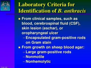
Results • Gram (+) Purple • Gram (-) Red • Difference • due to structure of cell wall • Gram (+) Thick cell wall • Gram (-) Thin cell wall A Gram stain of mixed Staphylococcus aureus
Ziehl–Neelsen stain • Differential Stain - divides bacteria into 2 groups • Acid Fast • Non Acid Fast • Used to identify organisms in the Genera Mycobacterium(high lipid and wax content in cell wall)
Procedure • Fix the smear of the specimen over the glass slide • either by heating or alcohol fixation • Pour carbol fuschin over smear • heat gently until fumes appear • do not overheat • allow it to stand for 5 minutes • wash it off with water
Pour 20% sulphuricacid • 5% sulfuric acid is used for destainingMycobacterium leprae instead of the 20% used for Mycobacterium tuberculosis • wait for one minute • keep on repeating this step until the slide appears light pink in color • wash off with water • Pour methylene blue • wait for two minutes • again wash with water • Allow it to air dry • examine under oil immersion lens
Result • Acid Fast organism • Red as Mycobacterium tuberculosis • Non Acid Fast organism • Blue as Enterobacteriaceae family A. Non Acid-fast bacteria B. Acid-fast bacteria Mycobacterium tuberculosis(stained red) in tissue (blue)
Special stains • Capsule stain and Flagella stain Pseudomonas fluorescensculturedon nutrient agar, stained usingthe Presque Isle flagella stain Encapsulated Bacillus sp. stained using Maneval'scapsule staining method
(II) Cultural Characters • Bacteria need nutritive culture media to multiply in vitro • An undefined medium (also known as a basal or complex medium). It is a medium that contains: • 1- A carbon source such as glucose for bacterial growth • 2- Water • 3- Various salts needed for bacterial growth • Defined media (also known as chemically defined media or synthetic media)
Classification of Media • Media can be classified into • 1-Minimal media ( simple medium) • It contains the basic nutritive requirements • e.g. nutrient broths and agar media
2- Selective media • Selective media are used for the growth of only selective microbes • It contains antibiotics, dye, or specific chemicals • inhibits the growth of most types of microbe • stimulate the isolation of one type
Mannitol salt agar (MSA) • selective for Gram positive (+ve) bacteria An MSA plate with Micrococcus sp. (1), Staphylococcusepidermis (2) and S. aureuscolonies (3).
Blood-free, charcoal-based selective medium agar (CSM) • isolation of Campylobacter sp. Blood-free, charcoal-based selective medium agar (CSM) for isolation of Campylobacter.
Löwenstein–Jensen medium • enriched selective media for T.B. Distinctive clusters of colorless Mycobacterium tuberculosis Löwenstein-Jensen mediumused for growing M. tuberculosis in a McCartney bottle
TCBS agar (Thiosulfate-citrate-bile salts-sucrose agar) • selective for Vibrio cholerae due to alkaline pH Yellow coloured (sucrose fermenting) colonies of Vibrio cholerae on TCBS agar.
3-Differential media • Differential media or indicator media • distinguish one microorganism type from another growing on the same media • Indicators • neutral red • phenol red • eosin Y • methylene blue
Examples of differential media include • Eosin methylene blue (EMB) • differential for lactose and sucrose fermentation E. coli on EMB agar
MacConkey (MCK) • differential for lactose fermentation A MacConkey agar plate with an active bacterial culture
4- Enriched media • Enriched media contain the nutrients required to support the growth of a wide variety of organisms • including some of the more fastidious ones • Blood agar • Is an enriched medium in which nutritionally rich whole blood supplements the basic nutrients • It contains 5-10% human or animal blood
It shows the type of haemolytic activity of bacteria (complete, partial or non-haemolytic) Complete Haemolysis of RBCs(Beta Haemolytic Streptococci) Partial Haemolysisof RBCs(Alpha HaemolyticStreptococci)
Chocolate agar (heated blood agar) • enriched with heat-treated blood (40-45°C). Comparison of two culture media types used to grow Neisseria gonorrhoeaebacteria
Lofflers serum media • Horse serum + glucose in a ratio 3:1 • It is used for cultivation of Corynebacteriumdiphtheriae
5- Transport media • Transport medium is a simple organic medium • maintain the viability of all organisms in the specimen • without altering their concentration • This type of medium mainly used for temporary storage of specimens • being transported to the laboratory for cultivation
Examples of transport media include • Thioglycollate broth for strict anaerobes Thioglycollate broth medium is recommended to isolate strict anaerobes should an anaerobic infection be suspected
The colonial appearance on culture media • Shape • The colonies may be small (pin-point) fimbriate, flat or convex • Colour • The colonies may be colorless or bacteria produce endopigments which give the colonies a characteresticcolour • Staph. aureusproduce golden yellow colonies • Staph. albusproduce white endopigment • Staph. citreusproduce a lemon yellow endopigment • The bacteria may produce exopigments • Pseudomonas aeruginosaproduce a green exopigments in the surrounding media
Antimicrobial Chemotherapy • An antibacterial agent is a compound or substance that kills or slows down the growth of bacteria • Antibiotic(s) has come to include a broader range of antimicrobial compounds, including anti-fungal and other compounds • It is produced by microbes and is harmful to other microbes, except viruses
These include • beta-lactam antibacterial • penicillin (produced by Penicilliumnotatum) • cephalosporin • Compounds that are still isolated from living organisms • Aminoglycosides • Other chemotherapeutic agents produced by chemical synthesis • Sulfonamides • Quinolones
Classification of Antibiotics • According to agent action • Antibacterial agents are divided into two broad groups based on their biological effect on microorganisms • bactericidal agents kill bacteria • bacteriostatic agents slow down or stall bacterial growth
Bactericidal antibiotics • Antibiotics that inhibit cell wall synthesis • Beta-lactam antibiotics • penicillin derivatives, and cephalosporins • Aminoglycosidic antibiotics are usually considered bactericidal • although they may be bacteriostatic with some organisms
Bacteriostatic antibiotics limit the growth of bacteria by interfering with • bacterial protein production • DNAreplication • Or other aspects of bacterial cellular metabolism • This group includes • Tetracyclines • Sulphonamides • Trimethoprim • Chloramphenicol • Macrolides
Antibiotic sensitivity test • Antibiotic sensitivity is a term used to describe the susceptibility of bacteria to antibiotics • Antibiotic susceptibility testing (AST) is usually carried out to determine which antibiotic will be most successful in treating a bacterial infection in vivo
Testing for antibiotic sensitivity is often done by the Kirby-Bauer method ( Disc-diffusion method) • Other methods to test antimicrobial susceptibility include the E-test (also based on antibiotic diffusion) • Agar and Broth dilution methods for Minimum Inhibitory Concentration determination
In Kirby-Bauer testing, white wafers containing antibiotics are placed on a plate of bacteria. Circles of poor bacterial growth surround some wafers indicating susceptibility to the antibiotic.
This is most commonly used in the setting of medicine, where a particular organism has been found to infect a patient, and the doctor treating the patient is seeking guidance on what concentration of antibiotic is suitable.
The Dilution Method • Serial dilutions of antibiotics are incorporated in agar containing or broth culture media • The lowest concentration of antibiotic that prevents visible growth after an 18-24 hours incubation period is known as minimal inhibitory concentration (MIC)
The minimal bactericidal concentration (MBC) may be determined in broth dilution tests by subculturing the containers that show no growth on to antibiotic-free agar containing media • The lowest concentration of antibiotic that totally suppresses growth after overnight incubation is known as MBC