Download
1 / 24
240 likes | 289 Vues
Learn the step-by-step analysis of protein complexes using BN/SDS-PAGE, from cell fractionation to complex resolution. Explore the impact of detergents, membrane solubilization optimization, and various detection methods for complex identification.

E N D
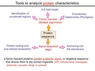
How to analyze protein complexes by 2D BN/SDS-PAGE Veronika Reisinger and Lutz A. Eichacker Department for Biology I, Menzingerstr. 67, 80638 München Reisinger/Eichacker Analysis BN-PAGE
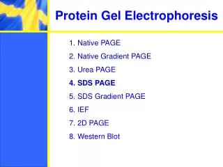
Workflow Cell fractionation Membrane isolation Solubilization of protein complexes BN-PAGE Denaturation of protein complexes SDS-PAGE Reisinger/Eichacker Analysis BN-PAGE
Detergents gal glu xyl Digitonin Dodecyl-ß-D-maltoside C56H92O29, Mr= 1229.31 g/mol C24H46O11 , Mr= 510.62 g/mol Reisinger/Eichacker Analysis BN-PAGE
Lhcb1 Lhcb1 Lhcb2 Lhcb2 Lhcb3 Lhcb3 PsbR PsbR PsbS PsbS psbC psbC psbD psbD psbA psbA Lhcb5 Lhcb5 Lhcb4 Lhcb4 Lhcb6 Lhcb6 psbB psbB psbE psbE psbI psbI psbF psbF psbK psbK psbH psbH PsbW PsbW psbM psbM psbN psbN psbL psbL psbJ psbJ Psb psb T T PsbO PsbO Psb Psb Q Q psbA psbD psbD psbA psbC psbC PsbP PsbP psbE psbF psbE psbF PsbO PsbO Membrane solubilization by Digitonin ( ) and DM ( ) H H M M Pet H Pet H Pet Pet H H E E Psa Psa 10kDa 10kDa Psa Psa psa psa C C Psa Psa D D Psa Psa G G Psa Psa Pet F Pet F Pet F Pet F PsbR PsbR 17,5 kDa 17,5 kDa 17,5 kDa psa psa A A psa psa B B J J I I 1 1 G G L L B B D D K K L L psa psa psa psa binding binding binding pet pet pet pet pet pet pet pet Psa Psa Psa Psa LhcA2 LhcA2 LhcA3 LhcA3 LhcA1 LhcA1 LhcA4 LhcA4 A A - - - FNR FNR FNR Psa Psa F F Psa Psa N N Pet Pet C C pet pet A A Pet Pet E E Pet E Pet E Below CMC, maintenance of membrane Above CMC, solubilization Reisinger/Eichacker Analysis BN-PAGE
Effect of detergent concentration on membrane solubilization 4.5-9 mM ß-DM or 9-18 mM Digitonin Decreasing molecular mass of protein complexes 1.1 mM ß-DM or 4.5 mM Digitonin Increasing concentration of detergent micelles Reisinger/Eichacker Analysis BN-PAGE
too low right too high too low right too high C D Digitonin (mM) ß-DM (mM) 1.1 2.2 4.5 9.0 18 144 9.0 36 36 72 144 1.1 2.2 4.5 18 72 kDa 669 669 440 440 232 232 Coomassie stained protein complexes 140 140 67 detergent concentration detergent concentration 67 too low too high too low right too high right Optimization of complex solubilzation A B ß-DM (mM) Digitonin (mM) 1.1 2.2 4.5 9.0 18 36 72 144 1.1 2.2 4.5 9.0 18 36 72 144 kDa kDa 669 669 440 440 232 232 Unstained protein complexes 140 140 67 67 detergent concentration detergent concentration Reisinger/Eichacker Analysis BN-PAGE
Optimized solubilization resolved by2D BN/SDS-PAGE B ß-DM (mM) Digitonin (mM) A BN-PAGE BN-PAGE 9.0 18.0 4.5 9.0 SDS-PAGE SDS-PAGE Reisinger/Eichacker Analysis BN-PAGE
Resolution analysis of protein complexes after2D BN/SDS-PAGE Separation of protein complexes of the thylakoid membrane by BN/SDS-PAGE II2 Subunit composition corresponding to one complex ? II1 Rubisco IIRC47 I/L1-4 I/L1 V IV BN-PAGE Pet H Pet H Psa G Psa D psa C Lhcb1 Lhcb2 psbC Lhcb5 Lhcb6 Lhcb4 psa B psa A psbD psbA I psa psbB G B D psbE psbF 1 1 psbI pet pet pet PsbW psbH psbK psbN psbL psbJ A - PsbO C Pet ATP synthase (IV) photosystem I (I/LI) cytochrome b6f (V) photosystem II (II) SDS-PAGE Schematic complex composition of the thylakoid membrane Reisinger/Eichacker Analysis BN-PAGE
Use of different detection methods after BN-PAGE or BN/SDS-PAGE approaches Reisinger/Eichacker Analysis BN-PAGE
Detection of protein complexes after BN-PAGE Coomassie staining Immuno detection Acitivity stain BN-PAGE Coomassie detection BN-Gelblot Immuno detection Marker BN-PAGE Marker BN-PAGE BN-PAGE Enzyme detection BN-PAGE 669 complexes exhibiting diaphorase activity 440 232 140 66 Reisinger/Eichacker Analysis BN-PAGE
Methods for the detection of proteins after 2D BN/SDS-PAGE : Post electrophoretic staining Methods for the detection of proteins after 2D BN/SDS-PAGE BN-PAGE BN-PAGE BN-PAGE SDS-PAGE SDS-PAGE SDS-PAGE BN-PAGE post electrophoretic staining DIGE BN-PAGE immuno detection BN-PAGE radioactive labelling SDS- PAGE 0min SDS-PAGE 20 min 80 min Reisinger/Eichacker Analysis BN-PAGE
In-gel staining Reisinger/Eichacker Analysis BN-PAGE
In-gel Coomassie staining of second dimension gels Reisinger/Eichacker Analysis BN-PAGE
Comparison of different staining methods after BN/SDS-PAGE : one gel, three stains Fluorescence Coomassie Silver Reisinger/Eichacker Analysis BN-PAGE
Methods for the detection of proteins:2D BN/SDS-DIGE BN-PAGE BN-PAGE SDS-PAGE SDS-PAGE BN-PAGE post electrophoretic staining DIGE immuno detection BN-PAGE SDS- PAGE BN-PAGE radioactive labelling 0min SDS-PAGE 20 min 80 min Reisinger/Eichacker Analysis BN-PAGE
CyDye labeling of membrane protein complexes Cy2 Cy3 Cy5 Ex Em 488 nm 532 nm 633 nm Reisinger/Eichacker Analysis BN-PAGE
2D BN/SDS-DIGE of plastid membrane complexes BN-PAGE BN-PAGE SDS-PAGE SDS-PAGE BN-PAGE condition I condition II SDS-PAGE condition I versus condition II + = Reisinger/Eichacker Analysis BN-PAGE
Methods for the detection of proteins after 2D BN/SDS-PAGE : Immuno detection BN-PAGE BN-PAGE SDS-PAGE SDS-PAGE BN-PAGE post electrophoretic staining DIGE immuno detection BN-PAGE SDS- PAGE BN-PAGE Radioactive labelling 0min SDS-PAGE 20 min 80 min Reisinger/Eichacker Analysis BN-PAGE
Detection of proteins in the native and denatured state BN-PAGE BN-PAGE SDS- PAGE No signal Not all proteins are detected by antibodies in the native state after BN-PAGE Good signal But in the denatured state after 2D BN/SDS-PAGE the same proteins are well detected Reisinger/Eichacker Analysis BN-PAGE
Detection of protein assembly kinetics by antibody detection after 2D BN/SDS-PAGE BN-PAGE timepoint 0 1 SDS-PAGE 2 3 Protein complexes Reisinger/Eichacker Analysis BN-PAGE
Methods for the detection of proteins after 2D BN/SDS-PAGE : radioactive labelling BN-PAGE BN-PAGE SDS-PAGE SDS-PAGE BN-PAGE post electrophoretic staining DIGE immuno detection BN-PAGE SDS- PAGE BN-PAGE radioactive labelling 0 min SDS-PAGE 20 min 80 min Reisinger/Eichacker Analysis BN-PAGE
Analysis of assembly by 2D BN/SDS-PAGE: Kinetic of protein complex formation followed by pulse/chase radiolabeling of de novo expressed proteins 0 min 80 min 20 min BN-PAGE SDS-PAGE Protein complexes Reisinger/Eichacker Analysis BN-PAGE
Summary for:The assembly analysis of protein complexesafter 2D BN/SDS-PAGEProtein subunits are identified by antibodies and/or mass spectrometry,andare labeled by fluorescent dyes and/or radioisotopes. Assembly is concluded from the position of protein subunits in the 2D -gel:A change of the horizontal position of a protein subunit from low to high molecular mass reflects the progression of subunit assembly into the corresponding protein complex Reisinger/Eichacker Analysis BN-PAGE
Step-wise assembly of proteins to complexes after 2D BN/SDS-PAGE: Protein subunits reveal the molecular mass increase of the complex M A T Pre Complex A Subunits N Complex R G A A A A A M M M Ü Ü A Ü Ü Ü Ü Ü T T T T T A N N N N T T T T T T T R R R D N N N N N N G G R R R R I G G G G G G G 6. A E E E 5. Direction of assembly 4. 1. 3. 2. 1. Native PAGE 2. Denaturing PAGE Reisinger/Eichacker Analysis BN-PAGE