Download
1 / 78
780 likes | 975 Vues
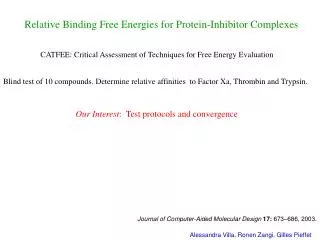
Relative Binding Free Energies for Protein-Inhibitor Complexes. CATFEE: Critical Assessment of Techniques for Free Energy Evaluation. Blind test of 10 compounds. Determine relative affinities to Factor Xa, Thrombin and Trypsin. Our Interest : Test protocols and convergence.

E N D
Relative Binding Free Energies for Protein-Inhibitor Complexes CATFEE: Critical Assessment of Techniques for Free Energy Evaluation Blind test of 10 compounds. Determine relative affinities to Factor Xa, Thrombin and Trypsin. Our Interest: Test protocols and convergence Journal of Computer-Aided Molecular Design 17: 673–686, 2003. Alessandra Villa, Ronen Zangi, Gilles Pieffet
Inhibitors 8 7 Inhibitor 2 10 9 6 1 3 4 5
Trypsin Template 2,6-diphenoxypyridine ligand PDB entry 1QB1 Acta Cryst. D (1999) D55 1395-1404
Factor Xa Template Removed from simulations 2,6-diphenoxypyridine ligand PDB entry 1FJS Biochemistry (2000) 39, 12534-42
Methods to Compute Free Energies Integration Formula (work along reversible path) Simulate at fixed and integrate numerically • Requirements (at each ) • Equilibrium. • Ensemble average must converge. • F() must be a smooth function. 1 2 3 4
Soft core potential: Addition of distance to shift the potential Alessandra Villa, Ronen Zangi, Gilles Pieffet
I3 to I4 In principle each point is independent I3 to I6 Alessandra Villa, Ronen Zangi, Gilles Pieffet
Cycle Closure: water (kJ/mol) -2.1 cycles 6 10 9 H H 2.8 C l N N N N N N C H 52.6 H 3 -321.5 -65.9 61.0 -28.6 -0.5 3.4 H H N 268.4 N H -97.9 O H N C H C H C H O O C H 3 3 3 3 + N 7 N 83.0 H C H 3 H 3 2 1 2 - - -3.4 -141.8 O O O O C H C H O O 3 2 238.8 -277.8 H 9.7 N N Cl N H -0.9 8 4 O C H C H O 3 2 Alessandra Villa, Ronen Zangi, Gilles Pieffet
Cycle Closure: Factor Xa (kJ/mol) 3.2 cycles 6 10 9 H H 2.1 C l N N N N N N C H 57.9 H 3 -304.3 -71.2 -36.5 6.0 72.3 -5.5 H H N 252.4 N H -97.9 O H N C H C H C H O O C H 3 3 3 3 + N 7 N 74.2 H C H 3 H 3 2 1 2 - - 12.1 -127.2 O O O O C H C H O O 3 2 245.7 -281.9 H 17.4 N N Cl N H 5.3 8 4 O C H C H O 3 2
Asymmetric Ligands + 1, 1* + hindered rotation in protein 4, 4* 5, 5* - Treat rotational isomers as independent compounds 6, 6* +
Asymmetric Ligands: Factor Xa 6* 10 9 -307.3 6 ? 3* 1* 269.5 -56.9 57.9 -304.3 3 1 2 7 Preferred conformers in factor Xa 252.4 74.2 4 5 5* 8 Alessandra Villa, Ronen Zangi, Gilles Pieffet
Comparison to Experiment G = Gfactor Xa - Gwater Ranking 1 >> 4 ~ 2 > 6 ~ 8 > 15 > 9 > 10 >> 7 Experimental data on the compounds investigated not yet released!. • Summary • Converged and reproducible results for mutations > 10 atoms(soft core potentials). • Large numbers of mutations possible (cluster computing). • Initial screening? (extrapolation approaches)
Estimating changes in the monomer-dimer equilibrium of SUC1 upon mutation Dissociation free-energy calculated using thermodynamic integration Gilles Pieffet
Protein of 113 residues a and b fold 4 stranded b sheets 3 short a helices Domain swapping of the dimer: C-terminal b strand SUC1: Structure Crystallographic structure of the monomer Crystallographic structure of the dimer Gilles Pieffet
Experiment Kd DM + M DGdiss = - RT ln Kd DDGDM = - RT ln ( ) mutant Kd WT Kd = DGdiss (M) - DGdiss (WT) Look at difference in disassociation constant upon mutation (experimental data not without question) Gilles Pieffet
Thermodynamic cycle DGdiss (WT) D (WT) M (WT) + M (WT) D (M) M (M) + M (M) DGWTM (dimer) DGWTM (mono) DGdiss (M) DDGDM = DGdiss (M) - DGdiss (WT) = - DGWTM(dimer) + 2 DGWTM(mono) Gilles Pieffet
Simulation parameters • Gromos96 forcefield • time step of 2 fs • T = 323 K • T and P coupling • Twin range cut-off of 0.9 and 1.4 nm • Reaction-Field (e=78) • Equilibration: 100 ps with position restraint 10 ns without position restraint • Free-energy calculation: • relaxation: 100 ps for each lambda point • data collection: • 400 ps for the monomer • 200 ps for the dimer • 18 l points are used for the integration Gilles Pieffet
Mutations studied All mutations correspond to the transformation of a residue into an alanine. DDGDM < 0 Dimer of WT is more stable than dimer in mutant DDGDM > 0 Dimer of WT is less stable than dimer in mutant Gilles Pieffet
Monomer: DG = 11.6 kJ/mol Sim: DDG = -2.2 kJ/mol dimer: DG = 25.4 kJ/mol Exp: DDG = -2.4 kJ/mol Gilles Pieffet
Monomer: DG = 4.6 kJ/mol Sim: DDG = -18.9 kJ/mol dimer: DG = 28.1 kJ/mol Exp: DDG = -0.4 kJ/mol Gilles Pieffet
Monomer: DG = 1.5 kJ/mol Sim: DDG = -5.1 kJ/mol dimer: DG = 8.1 kJ/mol Exp: DDG = -2.7 kJ/mol Gilles Pieffet
Monomer: DG = -5.2 kJ/mol Sim: DDG = -27.5 kJ/mol dimer: DG = 17.1 kJ/mol Exp: DDG = -2.3 kJ/mol Gilles Pieffet
Results Gilles Pieffet
Relative stability of the wild type dimer with respect to some mutants (kJ/mol).
Case of the LA95 mutation Monomer Gilles Pieffet
Results Gilles Pieffet
Monomer: DG = 4.6 kJ/mol Sim: DDG = -18.9 kJ/mol dimer: DG = 28.1 kJ/mol Exp: DDG = -0.4 kJ/mol Gilles Pieffet
Monomer: DG = 0.4 kJ/mol Sim: DDG = -4.2 kJ/mol dimer: DG = 3.4 kJ/mol Exp: DDG = 0.4 kJ/mol Gilles Pieffet
Monomer: DG = -3.5 kJ/mol Sim: DDG = -31.2 kJ/mol dimer: DG = 24.2 kJ/mol Exp: DDG = 0.4 kJ/mol Gilles Pieffet
Divergence for specific l values: l = 0.40, 0.45, 0.50, 0.55 for the monomer l = 0.30, 0.40, 0.45, 0.50 for the dimer Gilles Pieffet
Results Gilles Pieffet
Conclusions • no simple mutations when it comes to proteins • sampling on a multi ns timescale needed to get convergence due to protein fluctuations. • Not possible to tell if sampling/force field/structural problems. Gilles Pieffet
Incorporating the effect of ionic strength in free-energy calculations using explicit ions Serena Donnini and Alessandra Villa Why worry? Different protocols: ignore ions (ions independent, water high dielectric) neutralize the system (neutral system is more natural) add lots of ions Also: physiological ionic strength 0.1-0.2 molar. different claims concerning the creation of a net charge (create counter charge?) Generally ambiguous.
Incorporation of explicit ions in free energy calculations Cancellation of effects within a thermodynamic cycle? Protein charge = +2 Ligand charge = -2 Should one incorporate ions in the unlighted state? Will effects cancel? Serena Donnini and Alessandra Villa
Incorporation of explicit ions in free energy calculations • Consider a very simple mutation: • Only a change in dipole. • No change in number or atoms or net charge • Atoms partly buried. Thermodynamic integration 18 values simulate at fixed integrate numerically • Consider the same mutation in: • a charged molecule. • a neutral molecule • Ionic environment • no ions • just enough to neutralize charged system • 0.04M ionic strength • 0.1M ionic strength • 0.2 M ionic strength Serena Donnini and Alessandra Villa
Mutation of 2-phosphoglycolic acid (PGA) to 3-phosphonopropanoic acid (3PP) (triosephosphate isomerase inhibitors) pH ~2.0 pH ~7.0 Serena Donnini and Alessandra Villa
Mutation 2-phosphoglycolic acid (PGA) to 3-phosphonopropanoic acid (3PP) Bonded parameters
What did I expect? neutral form not much effect + charged form 2- some difference to neutral + 2 Na+ similar to neutral form more ions less effect + Serena Donnini and Alessandra Villa
Internal terms irrelevant Reaction field of the solvent
Incorporation of explicit ions in free energy calculations Ionic Strength Uncharged Species Charged Species Free energies are in kJ mol-1. Ionic strengths are in M. Slight trend Serena Donnini and Alessandra Villa
Uncharged Species 200 ps sampling at each l value Charged Species 200 ps sampling at each l value Serena Donnini and Alessandra Villa
Incorporation of explicit ions in free energy calculations No. of ions > 1 in close proximity negligible effect. Serena Donnini and Alessandra Villa
Incorporation of explicit ions in free energy calculations Serena Donnini and Alessandra Villa
Incorporation of explicit ions in free energy calculations minimum distance closest ion Average lifetime each ion ~ 7ns need 100’s ns at each lambda value no ions in close proximity Serena Donnini and Alessandra Villa
Conclusions • Experimentally the effect of the ionic strength on free energy differences is not expected to be very large. • Close proximity of ions has major effect even for mutations that do not involve change in net charge. • Inclusion of explicit ions can lead to severe sampling problems. • Options: • No inclusion of ions and accept errors associated with an overall charged system. • Perform simulations at high ionic strength to ensure sampling of ionic distribution.


![Binding energies in DATA [MeV]](https://cdn2.slideserve.com/3921996/slide1-dt.jpg)