Advanced Volumetric Brain Studies: Techniques and Findings Using 1.5 Tesla GE MRI Scanner
This study explores advanced volumetric brain analysis, utilizing a 1.5 Tesla GE MRI scanner. Key techniques include rigid registration and mutual information alignment for image acquisition. Brain segmentation leverages expectation maximization to classify four tissue types: white matter, gray matter, cerebrospinal fluid, and facial regions. Manual segmentation denotes regions of interest like the amygdala, hippocampus, and superior temporal gyrus, with statistical analysis revealing critical findings such as ventricular enlargement, lobe volume reductions, and changes in the basal ganglia. Technical challenges in image quality and segmentation methods are also discussed.


Advanced Volumetric Brain Studies: Techniques and Findings Using 1.5 Tesla GE MRI Scanner
E N D
Presentation Transcript
Current Technology • Image Acquisition: • 1.5 Tesla GE scanner, 1 coronal SPGR, 1 axial Double Echo PD and T2w image. • Rigid Registration: • Mutual Information Alignment. • Brain Segmentation: • Expectation Minimization Segmentation in 4 tissue classes (White, Gray, CSF and Face). • Manual Segmentation of Regions Of Interests. • Statistical Analysis of the ROIs’ Volume.
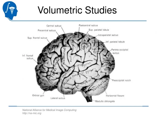
Brain Segmentation W. Wells
Posterior Superior Temporal Gyrus (STG)Red = Left, Green= Right• Amygdala. Yellow = Left, Light Blue = Right• Hippocampus (Hipp.). Orange = Left, Dark Blue = Right • Parahippocampal Gyrus (PHG). Dark Pink = Left, Purple = Right L. Post. STG L. STG Hipp. PHG L. Amygdala. L. Ant. STG L. Post. STG L. Hipp. Hirayasu et al.
Volumetric Studies Findings • Lateral Ventricles & CSF Enlargement. • Parietal Volume Reduction. • Temporal Lobe Volume Reductions • Amygdala-Hippocampal Complex, Parahippocampal Gyrus, & Superior Temporal Gyrus. • Prefrontal Volume Reduction. • Basal Ganglia (Caudate, Putamen, Globus Pallidus) increases and decreases.
Shape: Global Measures J. Levitt and C.F. Westin
Shape: Surface Based Analysis H.J. Park and J. Levitt
Shape: Medial Representations J. Levitt and S. Bouix
Technical Challenges • Image Quality and Acquisition Sequences. • Multi Channel Atlas Based Segmentation. • Multi Channel Image Registration. • Model Based Segmentation. • Shape Analysis. • Cortical Thickness.