Download
1 / 62
670 likes | 1.15k Vues
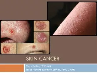
Skin Pre-Cancer and Cancer. Dr. Mary Cuthbert GPSI Dermatology. Sun, sea and sand…. There’s no such thing as a healthy tan. The effects of UV exposure -ageing of skin. -skin cancer. This presentation will cover :. Actinic keratosis Bowen’s disease Basal cell carcinoma

E N D
Skin Pre-Cancer and Cancer Dr. Mary Cuthbert GPSI Dermatology
This presentation will cover : • Actinic keratosis • Bowen’s disease • Basal cell carcinoma • Squamous cell carcinoma • Malignant melanoma • NICE guidance on skin cancer prevention
Actinic keratosis • Rough ,scaly spots on sun-damaged skin • Represent abnormal skin development due to exposure to UV radiation • Should be considered potentially precancerous(>10 AKs = 10-15% risk SCC) • Common on exposed sites eg backs of hands,face,scalp and ears of bald men
Actinic keratosis-treatment • Diclofenac gel (Solaraze) • Cryotherapy • Curettage/Excision • 5-Fluorouracil cream (Efudix) • Imiquimod 5% cream (Aldara) • Photodynamic therapy (not available in Bradford)
Bowen’s disease • Bowen’s disease is intraepidermal squamous cell carcinoma • It is effectively carcinoma-in situ • It may progress into squamous cell carcinoma (approximately 5%) • Because of this, it is very important to treat it effectively
Bowen’s disease • Presents as a pink or red ,irregular scaly patch • Usually develops in a sun –exposed area of skin • Common sites include hands and face in both sexes, scalp in men, lower legs in women • Diagnosis should be confirmed by biopsy
Bowen’s disease-causes: • UV radiation causes mutation in genes controlling skin cell growth • UV radiation suppresses immune response in skin • Arsenic ingestion • Ionising radiation-very common in early 20th century radiologists • HPV virus causes genital IEN
Bowen’s disease-treatment: • Cryotherapy • Curettage/excision • 5 Fluorouracil cream (Efudix) • Imiquimod 5% cream (Aldara) • Photodynamic therapy
Basal cell carcinoma • Affects fairskinned adults who have had a lot of sun exposure or repeated episodes of sunburn • Gorlin’s syndrome-inherited tendency to multiple BCCs • BCCs usually arise in normal-looking skin • BCCs grow slowly over months or years • Metastasis exceedingly rare but BCCs can cause destructive changes in surrounding tissues
Basal cell carcinoma-types: • Nodular BCC-most common type • Superficial BCC-common • Morphoeic BCC-waxy,scar-like • Pigmented BCC- can resemble melanoma • Basisquamous BCC-mixed BCC/SCC • Only the first two types are seen commonly in GP
Nodular BCC • Most common type on face • Small, shiny, skin-coloured swelling • Telangiectasia cross the edge • May have central ulcer or scab so edges appear rolled • Often bleed spontaneously, then heal over • Rodent ulcer is an open sore • Facial BCC should be referred to plastic surgeon
Superficial BCC • Often multiple • Upper trunk or shoulders commonest site but can appear anywhere • Pink or red scaly patch with raised edge on close examination • Slowly growing over months or years • Bleed or ulcerate easily
BCC- treatment: • Shave,curettage,cautery • Excision biopsy, may need grafting or flap. • Moh’s micrographic excision • Photodynamic therapy • Imiquimod 5% cream-highly effective for superficial BCCs • Cryotherapy • Radiotherapy
Squamous cell carcinoma • SCC is a common type of skin cancer • It develops in the epidermis from squamous cells which produce keratin • Usual presentation is a slowly –growing scaly or crusted lump • Can present as a non-healing sore or ulcer “punched out” in appearance • Sometimes growth is rapid over a matter of weeks
Squamous cell carcinoma-causes: • UV radiation-damages DNA in skin • SCC may develop in an actinic keratosis or patch of Bowen’s disease • Genetic predisposition to develop SCCs • Smoking-especially SCC lip • Thermal burns • Chronic leg ulcers • Immunosuppression-Azathioprine/Ciclosporin. Organ transplantation patients highly susceptible • HPV infection implicated in genital SCCs • Pre-existing skin conditions eg lichen sclerosus and lichen planus can predispose to development of genital and oral SCCs
Squamous cell carcinoma-treatment • If you suspect a possible SCC, refer via FAST TRACK pathway • Histological diagnosis confirmed in Dermatology department • Joint dermatologist/plastic surgeon assessment ideal, as happens in Bradford. • Specialist Skin Cancer Nurse input helpful • Surgery, possibly with skin graft • Radiotherapy may be needed
Metastatic Squamous cell carcinoma • 5% SCCs metastasise, most commonly from primary lesion on ear or lip • Commoner in transplant patients • Patients with CLL • Associated with increasing age • Associated with alcoholism • More likely if multiple skin cancers present
Malignant melanoma • Melanocytes are found in the basal layers of the epithelium • Non-cancerous growth of melanocytes results in moles or freckles • Cancerous growth of melanocytes results in malignant melanoma
Malignant melanoma-risk factors: • Sun exposure, particularly during childhood • Fair skin which burns easily • Blistering sunburn, especially when young • Previous melanoma • Family history of melanoma • Previous non-melanoma skin cancer • Large numbers of moles/ dysplastic moles
Common sites for melanoma: • In men commonest site is the back • In women commonest site is the leg • Can occur on mucous membranes, eg lips or genitals • Can occur under the nail • Can occur in eye, brain or mouth • BEWARE AMELANOTIC MELANOMA
MAJOR FEATURES: Change in size Irregular shape Irregular colour MINOR FEATURES: Diameter > 7mm Inflammation Oozing Change in sensation Glasgow 7 point checklist:
The ABCDE of melanoma • A Asymmetry • B Border irregularity • C Colour variation • D Diameter over 6mm • E Evolving (enlarging or changing)
Growth of melanomas • Horizontal growth within epidermis=melanoma in situ • Vertical growth through basement membrane into dermis=invasive melanoma • Once melanoma penetrates dermis,it spreads via lymphatic and blood stream = metastatic melanoma
Histological classification: Breslow thickness: • This is the thickness of the melanoma in mm Clark’s level: • This describes which layer of skin has been breached • Clark’s level 1-epidermis-melanoma in situ • Clark’s level 2-dermal invasion • Clark’s level 5- invasion of subcutaneous fat
Treatment of melanoma • Refer suspected melanoma via FAST-TRACK pathway • Surgical excision by Dermatologist with 2-3 mm margin • Wider excision if histology confirms melanoma • Thicker melanomas> 1mm-wider excision +/- sentinel node biopsy • Widespread melanoma-surgery/chemotherapy
Prognosis of melanoma • Breslow thickness< 1mm, almost 100% 5 year survival • Breslow thickness > 4mm, only 50% 5 year survival Remember, melanoma is a major cause of death from malignancy in young people
How can we advise our patients regarding skin cancer prevention?