Primary Active Transport
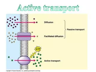
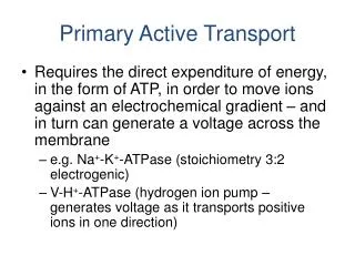
Primary Active Transport. Requires the direct expenditure of energy, in the form of ATP, in order to move ions against an electrochemical gradient – and in turn can generate a voltage across the membrane e.g. Na + -K + -ATPase (stoichiometry 3:2 electrogenic)

Primary Active Transport
E N D
Presentation Transcript
Primary Active Transport • Requires the direct expenditure of energy, in the form of ATP, in order to move ions against an electrochemical gradient – and in turn can generate a voltage across the membrane • e.g. Na+-K+-ATPase (stoichiometry 3:2 electrogenic) • V-H+-ATPase (hydrogen ion pump – generates voltage as it transports positive ions in one direction)
Primary Active Transport ATP → ADP + Pi 3+ ions out only 2+ in Vm -ve
Primary Active Transport • The ionic gradients are maintained by a combination of energy expenditure and Vm • Since the inwardly directed Na+ gradient is actively set up by the Na+-K+-ATPase – this acts as a membrane energy source, i.e. there is a large electrochemical gradient for Na+ to flow into the cell down an energy gradient – some transporters can use this Na+ gradient to transport other substrates (secondary active transport)
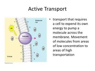
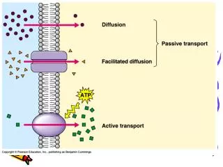
Secondary Active Transport. In this case an ion concentration gradient is used as the driving force to move a solute against its electrochemical gradient. The transport protein has a binding site for the ion that moves down its concentration gradient (in many cases Na+) as well as for the actively transported solute. The active movement may be in the same direction as that of the driving force ion (co-transporter/symport) or in the opposite direction (counter transport/exchangers).
Secondary Active Transporters • Do not rely on the hydrolysis of ATP directly • Co-transport – both substrates transported in same direction (generally the substrate is driven into the cell up a chemical gradient`uphill` – driven by the energy of Na+ flow into the cells down its electrochemical gradient)
Secondary Active Transporte.g. Na+/Glucose co-transporter [Na+] 145 Glucose gradient -60 mV [Na+] 15
1 Na+ binds to carrier. 3 Glucose binding changes carrier conformation. Intracellular fluid Lumen of intestine or kidney Na+ Na+ SGLT protein Glu Glu [Na+] high [Glucose] low [Na+] low [Glucose] high Na+ released into cytosol. Glucose follows. 4 2 Na+ binding creates a site for glucose. Na+ Na+ Glu Glu Secondary Active Transport
Exchangers • e.g. Na+/Ca2+ exchanger - the energy of the inwardly directed Na+ gradient drives Ca2+ out of the cell up its electrochemical gradient • In general exchangers utilize the electrochemical gradient of one substrate to drive another substrate in the opposite direction and generally up its electrochemical gradient
Endocytosis and Exocytosis Endocytosis is when regions of the plasma membrane engulf part of the extracellular fluid, pinch off and release their contents into the cell. Exocytosisis when a membrane-bound vesicle within the cell moves to the plasma membrane, fuses with it, and releases it contents into the extracellular fluid.
Receptor Mediated Endocytosis • Receptors on the plasma membrane detect and attach to the macromolecule • The receptors are concentrated in a region of the plasma membrane – clathrin coated pits • The pits bud from the membrane and eventually form a vesicle that is internalized • e.g. uptake of cholesterol by cells (low density lipoproteins bind to the LDL receptor and are taken into the cell in this way).
1 Extracellular fluid Ligand binds to membrane receptor. 9 Exocytosis Receptor-ligand migrates to clathrin-coated pit. 2 8 Transport vesicle and cell membrane fuse (membrane recycling). Clathrin- coated pit 3 Endocytosis Receptor Clathrin Transport vesicle with receptors moves to the cell membrane. 7 4 Vesicle loses clathrin coat. 4 Receptors and ligands separate. 5 To lysosome or Golgi complex Intracellular fluid 6 Ligands go to lysosomes or Golgi for processing. Endosome Receptor-Mediated Endocytosis and Exocytosis Exocytosis is the opposite of endocytosis
Low-Density Lipoprotein (LDL) Receptor Cells take up cholesterol by receptor-mediated endocytosis. Cholesterol is insoluble in water. Thus it is carried in tiny droplets of lipoprotein. The most abundant cholesterol carriers in humans are the low-density lipoproteins or LDLs. The first step in acquiring LDL particles is for them to bind to LDL receptors exposed at the cell surface. These transmembraneprotiens have a site that recognizes and binds to the apolipoprotein B-100 on the surface of the LDL. The portion of the plasma membrane with bound LDL is internalized by endocytosis People who inherit two defective (mutant) genes for the LDL receptor have receptors that function poorly or not at all. This creates excessively high levels of LDL in their blood and predisposes them to Atherosclerosis and heart attacks. The ailment is called familial (because it is inherited) hypercholesterolemia.
Epithelial Transport • This is the transport across the cells that line hollow organs or tubes. • This is important as they usually form a barrier between the lumen of the organ/tube and the blood. • Therefore the solute must be transported across two membranes in order to get to the blood from the lumen or vice versa. • The apical membrane is the cellular surface in contact with the luminal space (the outside), the basolateral membrane is in contact with the interstitium and ultimately blood. • Different transporters and pumps are present on each membrane, giving rise to a difference between membranes (polarity). • Membrane polarity allows for the directional transport of substances across epithelia.
Osmosis • Is a passive flow of the solvent • Net movement of a solvent across a partially permeable membrane from a region of high solvent potential to an area of low solvent potential down a concentration gradient. • Net movement of solvent is from the less-concentrated (hypotonic), to the more-concentrated (hypertonic) solution, which tends to reduce the difference in concentrations. • Osmotic pressure is the pressure that is applied to a solution to prevent such a flow.
Osmosis and Osmotic Pressure Osmolarity describes the number of particles in solution Figure 5-29
Osmotic pressure gradient is created by the presence of different concentrations of solutes in the solutions on either side of membrane. • Conc. Of osmotically active particles is expressed in osmoles (osm). • Osmolarity is the no. of osmoles per litre of solution eg. Plasma, • Osmolality- no. of osmoles per kilogram of the solvent. • Osmolalityof normal human plasma- 290 mosm/L