Download
1 / 41
440 likes | 744 Vues
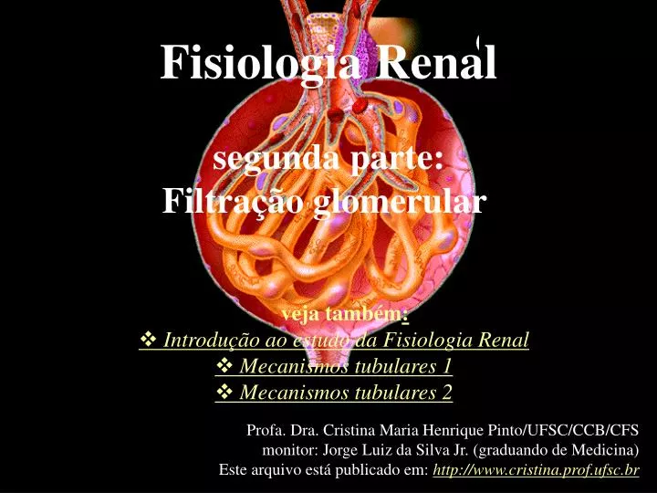
Fisiologia Renal segunda parte: Filtração glomerular. veja também : v Introdução ao estudo da Fisiologia Renal v Mecanismos tubulares 1 v Mecanismos tubulares 2. Profa. Dra. Cristina Maria Henrique Pinto/UFSC/CCB/CFS monitor: Jorge Luiz da Silva Jr. (graduando de Medicina)

E N D
Fisiologia Renal segunda parte: Filtração glomerular veja também: vIntrodução ao estudo da Fisiologia Renal vMecanismos tubulares 1 vMecanismos tubulares 2 Profa. Dra. Cristina Maria Henrique Pinto/UFSC/CCB/CFS monitor: Jorge Luiz da Silva Jr. (graduando de Medicina) Este arquivo está publicado em: http://www.cristina.prof.ufsc.br
Atenção: Recomendamos o material a seguir apenas com o objetivo de divulgar materiais de qualidade e que estejam disponíveis gratuitamente. Profa. Cristina Maria Henrique Pinto CFS/CCB/UFSC Esta apresentação é uma coletânea de figuras e textos extraídos da coleção em CD-ROM, utilizada em nossas aulas. “InterActive Physiology”, da Benjamin Cummings.
Você pode também dar baixa dos resumos dos CD-ROM´s, não apenas de Renal mas de diversos outros assuntos de Fisiologia Humana. Arquivos em *.pdf e/ou *.doc, com textos e ilustrações. Selecione: “assignments” na seguinte home page:http://www.aw-bc.com/info/ip/ E escolha entre os seguintes assuntos: Muscular; Nervous I; Nervous II; Cardiovascular; Respiratory; Urinary ; Fluids & Electrolytes; Endocrine(new) Veja também aulas online (DEMO dos CD-ROM´s) sobre: Endocrine System Cardiovascular System Immune System
Glomerular Filtration Graphics are used with permission of Pearson Education Inc., publishing as Benjamin Cummings (http://www.aw-bc.com )
Introduction • Formation of urine by the kidney involves three main processes: 1. filtration 2. reabsorption 3. secretion • Duringfiltration large quantities of water and solutes pass through the filtration membrane from the blood into the glomerular capsule. Goals • To preview the three major processes of the kidney. • To understand the function of the filtration membrane. • To understand the composition of the glomerular filtrate. • To understand the forces that determine glomerular filtration rate. • To examine the regulation of glomerular filtration.
Formation of urine by the kidney involves three main processes: • Filtration.The formation of urine begins with the process of filtration. Fluid and small solutes are forced under pressure to flow from the glomerulus into the capsular space of the glomerular capsule. • Reabsorption.As this filtrate passes through the tubules, specific substances are reabsorbed back into the blood of the peritubular capillaries. • Secretion.In addition, some solutes are removed from the blood of the peritubular capillaries and secreted by the tubular cells into the filtrate to “fine tune” the composition of the blood. • The rest of this topic covers filtration, while the filtrate processing topics in this module will cover reabsorption and secretion.
This simplified diagram of a kidney nephron shows the afferent and efferent arterioles, glomerular capsule, capsular space, glomerulus, the beginning of the renal tubule, and peritubular capillaries. • Label the diagram and draw arrows to indicate the processes of filtration, reabsorption, and secretion:
Humans produce ~1L of urine/day. Urine has a pH of ~6.0 and contains water and by-products of metabolism (i.e. urea, NaCl, KCl, phosphates, etc). The volume and composition reflects the volume consumed. • There are 3 steps to urine production: • Glomerular filtration • Tubular reabsorption • Tubular secretion
The Filtration Process • A coffee maker provides an everyday example of filtration. The paper filter acts as the filtration membrane. It holds back the large coffee grounds, while letting water and small solutes like caffeine and flavor molecules pass through. • A force is required to drive this process. In a coffee maker, it's the force of gravity. • What is the filtration force in the glomerulus? Yes! The hydrostatic pressure of the blood...
The Filtration Membrane and Glomerular Filtration • Just as a filter keeps grounds out of your coffee, the glomerular filtration membrane keeps blood cells and proteins out of the urine passageways. • The filtration membrane is composed of three layers: 1. Fenestrated glomerular endothelium 2. Basement membrane 3. Filtration slits formed by pedicels of the podocytes • Label this diagram:
Glomerular Filtration • Glomerular filtration is the process of passing solution through a selective barrier that separates particles based on size and/or electrical properties. • The filtration membrane is made of fenestrated endothelium, basement membrane, and filtration slits by pedicels of podocytes in the Bowman's Capsule. • The filtrate is fluid that is collected in the capsular space, which contains water, Na+, K+, Cl-, urea, uric acid, creatinine, glucose, amino acids, etc.
Glomerular Filtration • Passage through the filtration membrane is limited not only on the basis of size but also by electrical properties. • Glomerular filtration is a process of bulk flow driven by the hydrostatic pressure of the blood. • Small molecules pass rapidly through the filtration membrane, while large proteins and blood cells are kept out of the capsular space. • If the filtration membrane is damaged, proteins will leak through and end up in the urine. • If the filtration membrane is very badly damaged, then blood cells will leak through. • Consequences of loss of protein in the urine: • Loss of albumin causes a decrease in osmotic pressure. Fluid will leak into the interstitial spaces and edema results. Eventually there will be a low circulatory volume and possibly shock. • Loss of blood clotting proteins can cause uncontrolled bleeding. • Loss of globulins and complement proteins make the individual prone to infection.
Glomerular Filtrate • The fluid and solutes collecting in the capsular space is called glomerular filtrate. • The concentration of each of these substances in the glomerular filtrate is similar to its concentration in plasma. • Record the composition of the glomerular filtrate here: amino acids chloride creatinine glucose organic molecules potassium sodium urea uric acid nitrogenous waste water ions
Damage to Filtration Membrane • How might the contents of the filtrate be altered if the filtration membrane is damaged or destroyed? List factors here: _____________________________________________________ _____________________________________________________ • Presence of protein in the urine is called proteinuria. • Presence of blood cells in the urine is called hematuria.
Forces Affecting Filtration • There are three forces affecting filtration at the glomerulus: 1. Hydrostatic Pressure within the Glomerular Capillaries (Ph) • The blood pressure in the glomerulus averages 60 millimeters of mercury. This unusually high capillary pressure is the result ofthe short, large diameter afferent arterioles conveying blood at high arterial pressure directly to the glomerularcapillaries. • The smaller diameter of the efferent arterioles leaving the glomerulus also helps maintain the pressure by restricting the outflow of blood. • However, the glomerular hydrostatic pressure is opposed by two forces that reduce the flow of fluid into the capsular space.
Forces Affecting Filtration • The algebraic sum of these three forces produces the net filtration pressure of 17 millimeters of mercury. Ph= 60 mmHgPc= 15 mmHg Po= 28 mmHg Net Filtration Pressure = 60 mm Hg - (15 mm Hg + 28 mm Hg) = 17 mm Hg
Forces Affecting Filtration Analogy • An analogy for this process would be a football player (#60) trying to run for a touchdown. However, his progress to the goal line is slowed by two opposing players (#15 and #28). The runner eventually reaches the goal, but not as easily as he would without the resistance. • Now let’s see what happens when a smaller player carries the ball. Click the football to start the process. Here the two opposing forces, players #15 and #28, add together to counteract a smaller, weaker player, #43, stopping his forward progress. This situation is analogous to glomerular hydrostatic pressure too low to overcome the countering forces and carry out filtration. This is referred to as “kidney shut-down” or acute renal failure. Serious consequences will occur unless increased pressure and filtration are resumed.
Glomerular Filtration Rate (GFR) • The total amount of filtrate formed by all the renal corpuscles in both kidneys per minute is called the glomerular filtration rate, or GFR. In normal kidneys, the 17 millimeters of mercury of net filtration pressure produces approximately 125 milliliters of filtrate per minute. This translates to about 180 liters in 24 hours--nearly enough to fill a 50-gallon barrel! Fortunately, 99% of this volume will be reabsorbed as the filtrate passes through the tubules. • The glomerular filtration rate is directly proportional to the net filtration pressure, so a fluctuation in any of the three pressures previously discussed will change the GFR. Prolonged changes in normal GFR will cause either too much or too little water and solutesto be removed from the blood.
REGULATION OF GLOMERULAR FILTRATION RATE (GFR) AUTOREGULATION OF GFR (INTRINSEC MECHANISMS): MYOGENIC AND TUBULOGLOMERULAR FEEDBACK EXTRINSEC REGULATION OF GFR: SYMPATHETIC NERVOUS SYSTEM CONTROL TUBULOGLOMERULAR FEEDBACK PARTICIPATION (RENIN SECRETION)
Normal Mild Exercise Relaxation Systemic Pressure State of Afferent Arteriole Glomerular Hydrostatic Pressure Net Filtration Rate Autoregulation of GFR • Fill out this diagram as you work through this page: • During normal conditions, systemic blood pressure registers approximately 120 millimeters of mercury; the diameter of the afferent arteriole is normal, as is the glomerular hydrostatic pressure. These conditions provide a normal glomerular filtration rate of 125 milliliters per minute. • When blood pressure fluctuates during normal daily activities, autoregulatory mechanisms of the kidney alter the diameter of the afferent arteriole, in order to maintain a relatively constant glomerular filtration rate. • During mild exercise, the systemic blood pressure increases to 140 millimeters of mercury. If the afferent arteriole remains at the normal diameter, the 17% increase in glomerular hydrostatic pressure will cause a similar increase in GFR [of 21 milliliters per minute, to 146 milliliters per minute]. If allowed to continue, this increase will quickly cause severe dehydration. To avoid extensive fluid loss, the autoregulation mechanism decreases the diameter of the afferent arteriole, decreasing the glomerular blood flow. The glomerular hydrostatic pressure and GFR return to normal, even though the mild exercise continues and the systemic blood pressure remains elevated. • Reducing the activity level returns the systemic blood pressure to 120 millimeters of mercury; the afferent arteriole dilates to maintain normal glomerular hydrostatic pressure and GFR. • During periods of relaxation, the systemic blood pressure may drop to 100 millimeters of mercury or lower. If the diameter of the afferent arteriole remains normal, blood flow into the glomerulus is reduced. This in turn causes a reduction of glomerular hydrostatic pressure and GFR. To avoid poor filtering of the blood, the afferent arteriole dilates further to increase blood flow and glomerular hydrostatic pressure. Autoregulation has normalized the GFR.
Autoregulation of GFR GFR autoregulation Mechanisms:MYOGENIC MECHANISM • One of the mechanisms that provides autoregulatory control over renal processes is the result of the inherent tendency of vascular smooth muscle cells to contract when stretched. This allows the diameter of afferent arterioles to respond to changes in blood pressure. • When blood pressure in an arteriole increases, the walls of the vessel automatically constrict, reducing blood flow through the arteriole. • Low blood pressure reduces this reflexive constriction, so the arteriole dilates and blood flow is increased. • In renal autoregulation, this is called the myogenic mechanism. • Stretching the arteriole wall causes a reflexive vasoconstriction. As long as pressure continues, the vessel will stay constricted. • When pressure against the vessel wall is reduced, the vessel dilates. Changes in blood pressure therefore directly affect the constriction or dilation of the arteriole, and so glomerular blood flow.
High BP in Afferent Arteriole Low BP in Afferent Arteriole Effect on Wall of Afferent Arteriole Effect on Blood Flow to Glomerulus Autoregulation of GFR GFR autoregulation Mechanisms:MYOGENIC MECHANISM • Fill out this diagram as you work through this page:
Myogenic Autoregulation of GFR: Exercising If GFR was not automatically regulated in the body, it would increase when exercising due to the increased systemic pressure and glomerular hydrostatic pressure. Thankfully, it is autoregulated by constricting the afferent arteriole, thus returning glomerular hydrostatic pressure to normal, and returning GFR to normal.
Myogenic Autoregulation of GFR: Exercising If GFR was not automatically regulated in the body, it would increase when exercising due to the increased systemic pressure and glomerular hydrostatic pressure. Thankfully, it is autoregulated by constricting the afferent arteriole, thus returning glomerular hydrostatic pressure to normal, and returning GFR to normal.
Myogenic Autoregulation of GFR: Relaxed When we are relaxed, our systemic pressure decreases. If there was no autoregulation, our afferent arterioles would remain the same diameter, thus decreasing glomerular hydrostatic pressure and reducing GFR. Blessed are we with autoregulation... Our system reacts by dilating the afferent arterioles, thus increasing hydrostatic pressure, and consequently, returning GFR to normal.
Myogenic Autoregulation of GFR: Relaxed When we are relaxed, our systemic pressure decreases. If there was no autoregulation, our afferent arterioles would remain the same diameter, thus decreasing glomerular hydrostatic pressure and reducing GFR. Blessed are we with autoregulation... Our system reacts by dilating the afferent arterioles, thus increasing hydrostatic pressure, and consequently, returning GFR to normal.
Autoregulation of GFR GFR autoregulation Mechanisms: TUBULOGLOMERULAR MECHANISM • A second regulatory mechanism is the sensitivity of the macula densa cells of the juxtaglomerular apparatus tothe filtrateosmolarity and/or the rate of filtrateflow in the terminal portion of the ascending loop of Henle.
Autoregulation of GFR GFR autoregulation Mechanisms: TUBULOGLOMERULAR MECHANISM In response to high osmolarity of fluid in the distal tubule (GFR), macula densa cells release chemicals called vasoconstrictors (adenosine) that decrease the diameter of the afferent arteriole (GFR). In response to low osmolarity of fluid in the distal tubule (GFR), the macula densa cells release less vasoconstrictor.
Autoregulation of GFR GFR autoregulation Mechanisms: TUBULOGLOMERULAR MECHANISM In response to high osmolarity of fluid in the distal tubule (GFR), macula densa cells release chemicals called vasoconstrictors (adenosine) that decrease the diameter of the afferent arteriole (GFR). In response to low osmolarity of fluid in the distal tubule (GFR), the macula densa cells release less vasoconstrictor.
Autoregulation of GFR:TUBULOGLOMERULAR MECHANISM • High Osmolarity and High Rate of Filtrate Flow in Tubule. • Label the diagram below to indicate what happens when there is a high filtrate osmolarity: • A higher than normal concentration of sodium and chloride ions and/or a high filtrate flow rate in the terminal portion of the ascending loop of Henle indicates reduced reabsorption. This condition can be due to a high rate of filtrate flow through the tubules, resulting from an increased GFR. • This high osmolarity stimulates the macula densa cells to release vasoconstrictor chemicals, which cause the afferent arteriole to constrict. • The result is a lower GFR, slower filtrate flow, and increased tubular reabsorption of sodium and chloride ions.
Autoregulation of GFR:TUBULOGLOMERULAR MECHANISM • Circle the appropriate highlighted word(s) in the chart below: High GFR ¯ High/Low Filtrate Flow Rate in the Tubules ¯ Increased/DecreasedReabsorption of Ions in the Tubules ¯ High/LowOsmolarity in the Tubules ¯ Macula Densa Cells Release/Do Not ReleaseVasoconstrictor ¯ Afferent Arteriole Dilates/Constricts ¯ GFR Increases/Decreases ¯ Filtration Flow Rate Increases/Decreases ¯ Increased/DecreasedReabsorption of Ions in the Tubules ¯ Higher/LowerOsmolarity in the Tubules
Autoregulation of GFR:TUBULOGLOMERULAR MECHANISM • Low Osmolarity and low Rate of Filtrate Flow in Tubule. • A low rate of filtrate flow and decreased levels of sodium and chlorideions in the terminal portion of the ascending loop of Henle is also sensed by the macula densa cells. This condition is usually the result of slow filtrate flow due to low blood pressure and low GFR. In response, these cells decrease secretion of the vasoconstrictor chemicals. This leads to dilation of the arteriole and so increases blood flow, glomerular hydrostatic pressure, and the GFR. Label the diagram below to indicate what happens when there is a low filtrate osmolarity :
Autoregulation of GFR:TUBULOGLOMERULAR MECHANISM • Circle the appropriate highlighted word(s) in the chart below: Low GFR ¯ High/Low Filtrate Flow Rate in the Tubules ¯ Increased/DecreasedReabsorption of Ions in the Tubules ¯ High/LowOsmolarity in the Tubules ¯ Macula Densa Cells Release/Do Not ReleaseVasoconstrictor ¯ Afferent Arteriole Dilates/Constricts ¯ GFR Increases/Decreases ¯ Filtration Flow Rate Increases/Decreases ¯ Increased/DecreasedReabsorption of ions in the tubules ¯ Higher/LowerOsmolarity in the Tubules
Extrinsic regulation of GFR: tubuloglomerular feedback participation When the macula densa cells release less vasoconstrictorto low osmolarity of fluid in the distal tubule when GFR it may be uneffective to restore the GFR...
Extrinsic regulation of GFR: tubuloglomerular feedback participation Then, the macula densa cells signal the juxtaglomerular (JG) cells to release RENIN to the blood. Renin is a proteolytic enzyme whose release leads to increased levels of angiotensin II. Release of renin indirectly affects blood pressure and, hence, renal blood flow. renin
Extrinsic regulation of GFR: tubuloglomerular feedback participation The release of renin indirectly affects blood pressure and, hence, renal blood flow. Renin is a proteolytic enzyme whose release leads to increased levels of angiotensin II. Figure below shows effects of this hormone. Angiotensin II also stimulates release of aldosterone and vasopressin.
Extrinsic regulation of GFR:Sympathetic Nervous System Control • • Sympathetic nerve fibers innervate all blood vessels of the kidney as an extrinsic regulation mechanism. During normal daily activity they have minimal influence.However, during periods of extreme stress or blood loss, sympathetic stimulation overrides the autoregulatory mechanisms of the kidney. • • Increased sympathetic discharge causes intense constriction of all renal blood vessels. Two important results occur: • The activity of the kidney is temporarily lessened or suspended in favor of shunting the blood to other vital organs • 2) The lower GFR reduces fluid loss, thus maintaining a higher blood volume and blood pressure for other vital functions. As you can see, renal function has almost stopped. • • Reduction in filtration cannot go on indefinitely, as waste productsand metabolic imbalances increase in the blood. Autoregulation mechanisms are now ineffective in preventing acute renal failure. • • When fluid is given intravenously, the blood volume increases. With increasing blood volume, notice the gradual rise in blood pressure, reduction in sympathetic discharge, and restoration of renal functioning.
Veja animação online sobre o aparelho juxtaglomerular em: http://www.wisc-online.com/objects/index.asp?objID=AP2204 do Wisconsin Online Resource Center Mas cuidado: há erro! O filtrado quando passa pela mácula densa está subindo pela porção espessa ascendente da Alça de Henle (da região medular em direção ao córtex) e não descendo, como na animação...
Summary • Glomerular filtrate is formed by filtration of water and small solutes through the filtration membrane. • Net filtration pressure is the glomerular hydrostatic pressure minus the opposing forces of capsular hydrostatic pressure and glomerular osmotic pressure. • Blood pressure and flow in the nephron is monitored and controlled by renal autoregulation mechanismsin order to maintain a relatively steady glomerular filtration rate. • During periods of severe blood loss, the sympathetic nervous system overrides the renal autoregulatory mechanismsto shunt the blood to other critical areas.
Fisiologia Renal continua na terceira parte: Mecanismos tubulares I Veja mais sobre Anatomia e Histologia renais em: Introdução e filtração glomerular Mecanismos tubulares II ou em sugestões de WEBsites didáticos Profa. Dra. Cristina Maria Henrique Pinto/UFSC/CCB/CFS monitor: Jorge Luiz da Silva Jr. (graduando de Medicina) Este arquivo está publicado em: http://www.cristina.prof.ufsc.br