Download
1 / 31
310 likes | 472 Vues
The Respiratory System: Breath deeply the air of knowledge. Chpt 23. Respiratory System. Function : supply body with oxygen and dispose of carbon dioxide. Requirements. pulmonary ventilation (movement of air into and out of lungs) external respiration (gas exchange between blood and lungs)

E N D
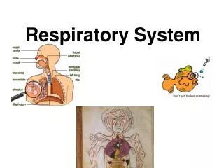
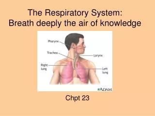
The Respiratory System:Breath deeply the air of knowledge Chpt 23
Respiratory System • Function: supply body with oxygen and dispose of carbon dioxide
Requirements • pulmonary ventilation (movement of air into and out of lungs) • external respiration (gas exchange between blood and lungs) • transport of respiratory gases (via blood) • internal respiration (gas exchange between blood and cells)
5 Functions of the Respiratory System • Provides extensive gas exchange surface area between air and circulating blood • Moves air to and from exchange surfaces of lungs • Protects respiratory surfaces from outside environment • Produces sounds-speaking, singing • Helps maintain pH
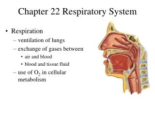
Anatomy of the Respiratory System Figure 23–1
Anatomy: Nose Functions • provides airway for respiration • moistens and warms entering air • filters air • serves as a resonating chamber for speech • houses olfactory receptors
Nasal Structures • External nose: bridge, root, dorsum nasi, apex, external nares, alae • Nasal cavity: nasal septum, internal nares (aka posterior nares or choanae), hard palate, soft palate, vestibule (with sebaceous and sweat glands and vibrissae) • Mucous membranes of the nasal cavity • olfactory mucosa (with smell receptors) • respiratory mucosa- secrete sticky mucus with antibacterial substances • Conchae- superior, middle, and inferior; help to trap particles
Sinuses • Paranasal sinuses • Frontal • Sphenoid • Ethmoid • Maxillary • Function to lighten skull & warm and moisten the air
The Nasopharynx • only an air passageway • during swallowing, closed off by the soft palate and uvula to prevent food from entering the nasal cavity • pseudostratified ciliated columnar • Pharyngeal tonsil (adenoids)
The Oropharynx • swallowed food and inhaled air • palatine & lingual tonsils • stratified squamous
The Laryngopharynx • Inferior portion of the pharynx • stratified squamous • where resp. and dig. pathways diverge
Larynx aka voice box • Functions • open airway • switching mechanism to route air and food into proper channels • voice production via vocal cords
Larynx aka voice box Anatomy • 9 cartilages connected by membranes and ligaments (8 are hyaline) • 9th cartilage is the epiglottis- elastic; closes over trachea during swallowing • Vocal folds aka true vocal cords- avascular; vibrate as air passes through them to produce sound • Medial opening between vocal folds is the glottis
Anatomy of the Trachea Figure 23–6
Trachea aka windpipe10-12 cm long; 2.5 cm diam • Layers of tracheal wall (internal to external) • Mucosa- pseudostr. ciliated columnar • submucosa • adventitia- with 16-20 rings of C-shaped hyaline cartilage, allowing flexibility
Trachea aka windpipe • 15–20 tracheal cartilages: • strengthen and protect airway • discontinuous where trachea contacts esophagus • Ends of each tracheal cartilage are connected by: • an elastic ligament and trachealis muscle • The carina (“keel”) marks the point where the trachea splits into the 2 primary bronchi (approx T7).
The Bronchial Tree • R and L primary bronchus • Secondary (lobar) bronchi- 3 R, 2 L • About 20 smaller branches • Bronchioles- less than 1 mm in diameter
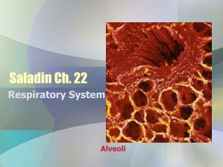
The Respiratory Zone: thin-walled alveoli (clustered into the alveolar sacs) where gas exchange occurs
Lungs • apex- narrow, superior tip • base- concave, inferior surface on diaphragm • L lung 2 lobes- upper and lower separated by oblique fissure • R lung 3 lobes- upper, middle, and lower separated by the horizontal and oblique fissures • Each lung lobe is divided into 10 bronchopulmonary segments
Relationship between Lungs and Heart Figure 23–8
Pleural Cavities and Pleural Membranes Figure 23–8
Pleural Cavities and Pleural Membranes • 2 pleural cavities: • are separated by the mediastinum • Each pleural cavity: • holds a lung • is lined with a serous membrane (the pleura)
The Pleura • Consists of 2 layers: • parietal pleura • visceral pleura • Pleural fluid: • lubricates space between 2 layers
Pleurae (membrane around the lungs) Parts of the parietal pleura. (parietal pleura in blue; visceral pleura in purple)
Grab a copy of the article: Struggling to Inhale • ANSWER THE FOLLOWING QUESTIONS • There are 2 different words for croup. List them and write what they each mean. • Explain how the virus that causes croup causes infection. • What is the treatment for croup? • What is a cricothyrotomy? Explain how doctors perform these. • What type of infection did the older patient have?