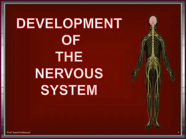
Development of the Nervous System
190 likes | 1.75k Vues
Development of the Nervous System . Development of the CNS. The embryo begins as a flat disk with three layers of cells. Endoderm forms the lining of the viscera (internal organs). Mesoderm becomes the bones and muscles. Ectoderm gives rise to the nervous system and skin.

Development of the Nervous System
E N D
Presentation Transcript
Development of the CNS • The embryo begins as a flat disk with three layers of cells. • Endoderm forms the lining of the viscera (internal organs). • Mesoderm becomes the bones and muscles. • Ectoderm gives rise to the nervous system and skin. • Neural plate becomes the nervous system.
Neurulation • At about 3 weeks, a rostral to caudal groove forms in the neural plate. • The two sides of this groove fold together and fuse forming a neural tube. • The entire nervous system develops from the neural tube. • Part of the tube pinches off and becomes the neural crest – the neurons of the peripheral nervous system.
Differentiation • Structures become more elaborate and specialized during development. • The neural tube forms three vesicles. • Forebrain becomes the neocortex. • Midbrain becomes the tegmentum and tectum (substantia nigra, inferior/superior colliculi) • Hindbrain becomes the cerebellum, pons, medulla, and brain stem.
Other Names for the Parts • Telencephalon and diencephalon – forebrain. • Mesencephalon – midbrain • Rhombencephalon -- hindbrain
Ventricles • Ventricles are open, fluid-filled spaces within the brain. • Cerebrospinal fluid (CSF) • Ventricles correspond to the three parts and can be used as landmarks across species. • Cerebral aqueduct (narrow channel) identifies midbrain. • Ventricles are continuous and continue into spinal cord.
Neocortex • The terms cortex and neocortex are used interchangeably when referring to humans. • Only mammals have neocortex. • Neocortex is used for different things in different species. • Evolution has its greatest impact on the size and function of the neocortex. • Localization of functions to areas of the brain is referenced to Brodmann areas.
Names for Collections of Neurons • Gray matter – neuronal cell bodies. • Cortex – a thin sheet of neurons on the brain’s surface. • Nucleus – a mass of neurons deep in brain. • Substantia – related neurons with less distinct borders than a nucleus. • Locus – small, well-defined group of cells. • Ganglion – group of peripheral NS neurons.
Names for Collections of Axons • Nerve – bundle of axons in peripheral NS. • White matter – CNS axons. • Tract – axons with a common origin and destination. • Bundle – axons that run together for awhile. • Capsule – axons connecting brain stem with cortex. • Commisure – axons connecting hemispheres. • Lemniscus – a tract that runs through the brain like a ribbon.