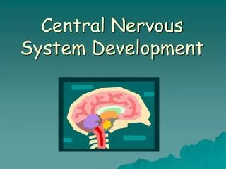
Development of the Nervous System
590 likes | 1.28k Vues
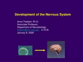
Development of the Nervous System. Anne Theibert, Ph.D. Associate Professor Department of Neurobiology theibert@nrc.uab.edu 4-7278 January 8, 2009. 3. 2. 1. Embryonic. 5. 6. 7. 8. 9. Fetal. 1. 15. B. 3. 6. 24.

Development of the Nervous System
E N D
Presentation Transcript
Development of the Nervous System Anne Theibert, Ph.D. Associate Professor Department of Neurobiology theibert@nrc.uab.edu 4-7278 January 8, 2009
3 2 1 Embryonic 5 6 7 8 9 Fetal 1 15 B 3 6 24 Development of Nervous System during Prenatal and Postnatal Periods Prenatally: Gross structures Most neurons 350-400 g Genetic factors Postnatally: Few neuronal populations Many glia Dendritic arborization Synaptogenesis Myelination 1200-1400 g Environment Experience
Stages of Development of the Nervous System • Initial Embryonic Development: Fertilization, cell divisions, implantation, and gastrulation 2. Neurulation:Establishment of primordial nervous system in early embryo • 3. Proliferation:Initial generation of neuronal/glial precursors from undifferentiated precursor cells 4. Migration/Aggregation:Movement of neurons and glia from the sites of generation to their final positions 5. Differentiation:Determination of the type of neuron or glial cell 6. Targeting/Synaptogenesis: Formation of axon pathways and synaptic connections 7. Programmed Cell Death/Synapse Refinement:Some neuronal death, elimination of some synaptic connections, changes in synaptic strength 0- 2 wks 2 wks - 5 wks 5 wks - 6 mos PN 5 wks - 25 wks 25 wks - 6 mos PN 25 wks - adult 25 wks - adult
Initial Embryonic Development Cleavage of fertilized egg multicelled blastocyst (inner cell mass and trophectoderm) (5 days) Cellular movements of Inner cell mass forms hypoblast and epiblast Epiblast is a two-layered disc of cuboidal cells-forms embryo Hypoblast cells adjacent to epiblast flatter cells- forms yolk sac Trophectoderm forms placenta Cleavage and Cell Division Fertilization trophectoderm trophectoderm
Gastrulation involves cell migration Rostral Caudal Epiblast Hypoblast Primitive Streak Wall of Yolk Sac Area of Prechordal Plate Gastrulation converts epiblast into three germ layers Epiblast cells move along surface pile up, then move internally Primitive streak: Dip in dorsal midline on ectoderm Becomes neural groove as cells move in Human Day 14-17
Gastrulation forms three primary germ layers Cells migrating first replace some hypoblast cells become the endoderm Cells move in later migrate over the endoderm form mesoderm. Cells that remain on surface form the ectoderm Epidermis and associated structures (skin, nails, hair) Nervous system (CNS and PNS) Muscle, circulatory system, bones and cartilage, outer covering of internal organs, excretory system, and gonads Inner lining of digestive system, inner lining of respiratory system, glands (including liver and pancreas)
Formation of Neural Plate and Notochord Ectoderm cells on dorsal side form neural plate Neural Plate becomes population of progenitor cells that gives rise to neurons and glial cells Neural Plate-broad rostrally at brain plate and narrow caudally at spinal cord plate Notochord Notochordal tissue separates from mesoderm below neural plate to form notochord Signals from notochord specify which ectoderm cells become neural plate Notochord is transient structure in mammals but helps specify neural tube differentiation
Neural Induction in vertebrates What are signals from the organizer? Secreted Factors: Noggin/Chordin/Follistatin How do they induce? Inhibitors of BMP signaling BMP: bone morphogenetic protein (members of TGFfamily of polypeptide growth factors) Amphibian BMPs expressed in ventral region, have strong ventralizing activity BMPs act on ectoderm to form epidermis. BMP activity inhibits neural fate. Block BMPs with N,C,F, ectoderm becomes neural. Neural fate may be default pathway
Process by which the neural tube is formed Neural tube is the precursor to the central nervous system The peripheral nervous system arises mainly from neural crest and placodes Occurs from 2 - 5 weeks (humans), sets up the major brain and spinal cord regions, before proliferation and differentiation begins-- Critical period during which errors can cause severe birth defects Neurulation Neurectoderm Neural groove Primary neurulation, neural plate folds to produce the neural tube. Cells at specialized regions of neural plate undergo shape changes causing plate to buckle inwards (neural groove) elevating the margins (neural folds).
Neural groove forms in the midline of the neural plate about day 18-19, continues to deepen until about day 24-27 Neurulation Shape of the neural plate changes dramatically as cells undergo an active rearrangement process. Movements of cells brings the neural folds together.
Neurulation 4th week Bending of the plate followed by neurulation Folds meet and fuse at dorsal midline--roof plate Pinch off from the surrounding ectoderm Forms neural tube that separates from non-neural ectoderm- becomes epidermis Tube larger at anterior---narrow at posterior Neural tube-- hollow Fusion of the neural folds requires cell adhesion molecules. Neural plate ectoderm expresses N-cadherin and N-CAM while epidermis expresses E-cadherin
Primary Neurulation Fusion of neural folds begins at 4th somite about 21 days Extends rostrally and caudally Neural tube fuses at different times along length Leaves two openings at either end-neuropores Head folds fuse before tail folds Primary phase forms the brain and rostral (anterior) spinal cord Secondary phase when the caudal (posterior) region of the spinal cord is formed. Anterior/Rostral neuropore normally closes around day 23-24 Caudal/Posterior neuropore closes about 2 days after the anterior neuropore (day 27), after the embryo has curved ventrally Failure of proper neural tube closure results in neural tube defects
Secondary Neurulation Caudal end of neural tube formed by secondary neurulation. Develops from the primitive streak region. Region called the mesodermal caudal eminance sinks under the epidermis, forming medullary cord, which then condenses and cavitates (this part of the neural tube not derived from ectoderm) •Beyond point of posterior neuropore •Days 20 to 42 •Joins and becomes continuous with neural tube
Neural Tube Defects • Most common neurological malformation in humans at birth. • Serious birth defects that involve incomplete development of the brain, spinal cord and/or protective coverings for these organs, can result in stillbirth or death shortly after delivery. • Failure of proper neural tube closure or bending result in the neural tube remaining open or not undergoing proper morphological changes. • Most common NTDs are Anencephaly and Spina Bifida Each occur in approximately 1-2/1000 births Folate Failure of anterior neural plate to fuse/close (region 2) Failure of posterior neuropore to fuse/close (region 5)
Segmentation of Neural Tube Constrictions subdivides rostral/anterior part into three vesicles --- primordia of the brain. First division forms of 3 vesicles form forebrain Prosencephalon midbrain Mesencephalon hindbrain Rhombencephalon, and Spinal Cord Vesicles expand because tube is filled with fluid Regional differences in extent of swelling caused by expansion of epithelium under pressure from fluid in the tube. Pressure requires transient blockage of neural tube between brain and spinal cord. Spinal Cord
4th wk Segmentation of Neural Tube Neural tube undergoes bending movements to become flexed. Earliest flexure in region of midbrain (cephalic flexure) results in the forebrain swinging posteriorly beneath the hindbrain. Second flexure appears caudally (cervical flexure) in region of hindbrain spinal cord junction
Segmentation of Neural Tube Two regions become divided into sub-regions (to produce Five Secondary Vesicles) Each develop into specialized region of brain Cerebrum:Cerebral Hemispheres-Neocortex/Basal Ganglia/Hippocampus Thalamus/ Hypothalamus/Retina/ Optic Nerve Midbrain: Tectum and Tegmentum-Colliculi and monoamine neurons Pons/Cerebellum Medulla Prosencephalon Rhombencephalon 5th wk
8-weeks Early morphological features of neural tube reflect overall plan of the CNS Predict later regional specialization. Neural tube is a simple epithelium-single cell thick. Germinal neuroepithelium serves as source of nearly all central neurons and glia.
Cavities within the tube form ventricles Ventricular System Lateral ventricles derived from the telencephelon Interventricular foramen Cavity of diencephalon forms Third ventricle Cavity of the midbrain forms Cerebral aqueduct Cavity of hindbrain forms Fourth Ventricle Telencephalon Diencephalon Mesencephalon Metencephalon Myelencephalon
Patterning along the rostral-caudal axis Transcription Factors Hox genes: Transcription factors that contain a homeobox domain (also called homeotic genes) that are expressed along the embryo rostral-caudal axis, regulate transcription of other developmentally regulated genes Midbrain/Hindbrain Boundary
Neural tube also patterned along its D/V axis D/V information conveyed to neural tube by dual signals from notochord and dorsal epidermis. Dorsal epidermis induce signaling center in roof plate. Dorsal signals from epidermis are BMPs (BMP4/7, dorsalin). Roof plate responds to BMPs by making more, sets up gradient. BMPs SHH Notochord produces SHH: Sonic Hedgehog protein induces floor plate to produce SHH SHH gradient allows differential expression of transcription factors along the D/V axis, determines fates of cells along the ventral/dorsal axis (V3, motor neurons, V2, V1).
Errors in Development Holoprosencephaly Lobes fail to separate to different degrees Alobar Holoprosencephaly accompanied by the failure of fetal facial midline structures to form properly. Usually midline facial defects (cleft lip, cleft palate, cyclopia, etc) accompanying this condition. Single large ventricle, because there was no separation of cerebral hemispheres. Usually babies do not survive beyond one year. • Some caused by mutations in SHH • 1 in 16,000 live births • fetal alcohol syndrome/maternal diabetes mellitus • Sporadic, 10% genetic (sonic hedgehog, trisomy 13 and 18) • alobar (63%), semilobar (28%), lobar (9%)
Separation of Sensory and Motor Neurons in Spinal Cord and Brain Stem D/V axis, tube is subdivided into two dorsal (left and right) alar plates, joined in midline by roof plate Two ventral basal plates united in midline by floor plate. Furrow- sulcus limitans forms between alar and basal plates about the 4th week.
Proliferation/neurogenesis 5 wks - 7 mos PN After neural tube closes, neuroepithelium is one cell thick. Long cells touch both basal lamina (outer surface of neuroectoderm) and the apical surface (luminal/ventricular). Proliferation (mitosis/cell division) of neural stem cells takes place and becomes the ventricular zone. Two different cleavages during mitosis. Vertical cleavage (get two identical stem cells). Horizontal cleavage (one cell remains connected to lumen and remains a stem cell and other cell will not divide and migrates away.
Proliferation/Neurogenesis Dividing precursor cells in the ventricular zone undergo stereotyped pattern cell movements as progress through mitotic cycle forming either new stem cells or postmitotic neuroblasts that differentiate into neurons or glia. Cells withdraw (undergo last S phase) at different times. Neuron’s birthday. • Ventricular zone progenitor cells start dividing (symmetrically) E28 • VZ asymmetric cell growth (neurogenesis) starts E42 • Greatest production of neurons E42-E125 • Brain forms from single layer of cells • Mature human brain contains 100 billion neurons • At peak, high rate of proliferation, 250,000 neurons produced per minute
Neurogenesis continues throughout life in humans Hippocampus (dentate gyrus), olfactory bulb Subventricular layer/zone
Migration in CNS Once produced, neurons migrate to what will be their permanent position in the brain, where they will connect with other cells to form the major parts of the brain Many neurons arrive at their final position by 5 months; faulty neural migration may lead to developmental disorders CNS, migrating cells not proliferating. At the same time the neurons are forming some cells serve as pathway for neurons that form the cerebral cortex (called-radial glia but many become neurons). New cells migrate outwardly towards the cortical surface. Each migration passes previously migrated cells.
Development of Layers of Cerebral Cortex I VZ: ventricular zone IZ: intermediate zone PP: preplate CP: cortical plate MZ: marginal zone II MZ III IV PP CP V IZ IZ VI VZ VZ VZ WM Neuronal Migration during Neurogenic Interval Young neurons migrate across the embryonic cerebral wall to the neocortex in an inside-to-outside order (layer VI develops first and layer I develops last). At the conclusion of migration a portion (20-50%) of neurons are eliminated through apoptosis (programmed cell death). The remaining neurons grow, differentiate, and become integrated into the cortical circuitry.
Signaling Molecules Reelin is secreted ECM protein required for corticogenesis ApoER2 and VLDLR are important signaling receptors that bind reelin and regulate neuronal migration in the developing brain, while megalin -deficient mice exhibit a classical defect of forebrain development, holoprosencephaly. Pial surface Cajal-Retzius neurons Migrating neuron Migrating neuron Radial Glial cell Ventricular zone Reelin VLDLR mDAB (adaptor) ApoER2 Megalin
Disorders of brain growth and migration Cortical heterotopia Displacement of grey matter into white matter Smooth brain-migration defect- no gyri or sulci Microcephaly Encephalocele (brain hernia) a rare defect (1 in 10,000-20,000 births) caused by a hole in the skull through which brain tissue protrudes (herniates) Condition generally results in death shortly after delivery Frontal Occipital Encephalocele (brain hernia)
PNS Development Majority of PNS neurons and glia derived mainly from Neural Crest cells which form at the dorsal aspect of the neural folds. Somatic division (primary motor and sensory neurons of the body) Autonomic division (sympathetic, parasympathetic, and enteric neurons) of nervous system Sensory epithelia of nose, inner ear, lens, anterior pituitary gland, and neurons of certain sensory ganglia of cranial nerves are derived from placodes, epithelial thickenings that form in otherwise epidermal ectoderm around the margins of the anterior neural plate
PNS Development Neural crest cells- dorsal to the neural tube Pluripotent cells that migrate away from the crest area and develop into PNS neurons & glia: sensory neurons (DRG), some cranial nerves, postganglionic neurons of autonomic nervous system, Schwann cells, satellite cells, melanocytes, adrenal chromaffin.Cranial neural crest components extend from the level of the diencephalon to the 5th somite. Cranial neural crest participates in formation of periocular tissues, and forms skeletal elements of the branchial arches.
Peripheral Nervous System Migration Guided by adhesion molecules (neural crest cells) •Wnt family of genes induces delamination, lose tight junctions and diminish adhesion molecules- cells become migratory •Travel through very specific route because it is a permissive environment and gives substrate (ECM) for migration (specific laminin, glycoproteins, proteoglycans). •When they reach their destination, they upregulate adhesion molecules •Proliferate as they migrate (different from CNS) •Intrinsic and extrinsic information determines fate Neural Crest
Differentiation/Determination Intrinsic and Extrinsic Cues Transcriptional activators or repressors Diffusible molecules, signals on membranes, ECM bound molecules
Target Selection Multiple cues in axon guidance Growth Cone Requires both Microtubule (tubulin) and microfilament (actin) - based cytoskeletal changes Signaling through surface receptors
Targeting/Synaptogenesis Synaptogenesis is process of synapse formation, rapid during early years, continues throughout life. Occurs at different rates for different parts of the brain. Visual system: peaks between 4 and 12 months, peak number of synapses in prefrontal cortex not until 24 months of age. Adult density of synapses for visual cortex is attained between 2 and 4 years; prefrontal cortex- not until teens; dendritic branching increases from infancy to adulthood. Whether synapses will be formed and maintained and whether neurons will live or die? Experience.
Synapse Elimination Imparts specificity Depends on activity
Normal DS MR FraX Spine and Synapse dysgenesis in mental retardation Neurodevelopmental disorders affecting wiring Down syndrome, mental retardation, Fragile X, Autism
Critical Periods of Development Teratogens Agents that cause congenital malformations during critical periods, and subtle alterations in the brain during sensitive periods