Download
1 / 63
650 likes | 1.2k Vues
OB/Gyn Grand Rounds: What Can the Interventionalist Do for Your Patients. Thea Moran, MD Asst Professor Louisiana State University New Orleans, LA. Disclosures. I have no disclosures to make. What can the IR do for the OB/Gyn service?. 1. Uterine fibroid embolization

E N D
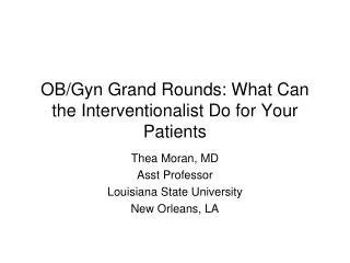
OB/Gyn Grand Rounds: What Can the Interventionalist Do for Your Patients Thea Moran, MD Asst Professor Louisiana State University New Orleans, LA
Disclosures • I have no disclosures to make
What can the IR do for the OB/Gyn service? • 1. Uterine fibroid embolization • 2. Nonfibroid uterine artery embolization • 3. Prepartum uterine artery embolization • 4. Pelvic congestion syndrome • 5. Fallopian tube recanalization • 6. Endovenous ablation of varicose veins
1. Uterine fibroid embolization • Fibroids, or leiomyomas, are benign smooth muscle tumors of the uterus occurring in reproductive age women. • Most common benign tumors of the female genital tract although many are without symptoms • Fibroids are most frequent in 30-40 year olds and involute after menopause • Goal of UFE is selective infarction of symptomatic fibroids
Symptoms • Menstrual abnormalities • Menorrhagia • Most frequent indication • Symptom most responsive • DDX: adenomyosis, endometrial polyps, endometrial hyperplasia, endometrial CA • Menometrorrhagia • Worsening of menstrual cramps • Pressure symptoms • Pressure sx related to the bladder, ureter, bowel, or nerves • DDX: ovarian and abdominal masses, infection, endometriosis, adenomyosis; fibroid torsion, degeneration or prolapse; PID, endometriosis, adenomyosis, pelvic congestion syndrome, GU/MSK/GI causes
Symptoms • Dyspareunia • Increasing pelvic or abdominal girth • Needed premyomectomy, hysterectomy for fibroids • Infertility or miscarriages with no other discernible cause • Controversial
Contraindications • Severe anaphylactoid reaction to contrast • Uncorrectable coagulopathy • Severe renal insufficiency • Pregnancy • Gynecological malignancy • Active pelvic infection/inflammatory disease • Prior pelvic radiation • Microvascular disease • Pedunculated or mucosal fibroids • Expulsion, cervical impaction
Uterine artery anatomy • First branch of anterior division of internal iliac artery • 3 segments • Descending segment goes along pelvic wall • Transverse segment goes to the midline • Ascending segment along the uterus
Uterine artery anatomy • Cervicovaginal artery • Perforating arteries, terminal branches to the fallopian tubes and ovaries • Inferior vesicle artery has common origin • Can have completely or partially absent uterine artery which can be bilateral
Ovarian artery anatomy • 1 ovarian artery per ovary • Originate from aorta a few cms below renal artery origins • 4% of women have ovaries supplied 100% by the uterine artery • Nikhil C Patel, Interventional Radiology Secrets.
Ovarian artery anatomy • 46% have uterine-ovarian artery anastomoses * • 5-10% have angio vis anastomoses * • Ovarian blush • Avoid nontarget embolization • Normal diameter: 1 mm • * Nikhil C Patel, Interventional Radiology Secrets
Preoperative work up • History, pelvic exam, informed consent • CBC, FSH (day 3), chem 7, PT/PTT/INR • Pelvic US • Usually for screening and sufficient • Uterine and fibroid volumes, fibroid position • Pelvic MRI • Better definition of fibroids, uterus c/w US, evaluate for adenomyosis and other pathology • CE MRA help planning of UFE • Demonstrates vascular supply to fibroids • PAP smear • R/o cervical CA
Preop work up • Endometrial biopsy • R/o endometrial cancer • US and MRI can’t definitively evaluate endometrial thickness • Intermenstrual or irregular bleeding, postmenopausal women with vaginal bleeding • All women over 40 yo with menorrhagia • D/c GnRH analogues several weeks preop • Fibroids enlarge
Patient consultation • Does the patient desire future childbearing? • Weigh treatment options • Severity of symptoms severe enough physically and/or psychologically to warrant invasive therapy especially since most fibroids involute after menopause
Treatment options • Medical • NSAIDS, OCP • Mild symptoms • GnRH • Moderate symptoms • Decreases estrogen which causes fibroid degeneration after 3 months of therapy • Noninvasive, short term relief of symptoms, good for perimenopausal women, adjuvant therapy preoperatively • Not to be given indefinitely • Surgical • Severe symptoms • Open, laparoscopic or hysteroscopic techniques • Hysterectomy • Complete cure, r/o possibility of future neoplasm, facilitates postmenopausal HRT, invasive
Treatments options • Myomectomy • Preserves uterus and future childbearing • Bleeding c/o if multiple fibroids • High recurrence rate (20-25%) • Nikhil C Patel, Interventional Radiology Secrets. • UFE • Severe symptoms • Uterus retained, all myoma treated at once, noninvasive • Postembolization syndrome, potential for contrast reaction, possibility of no symptom relieve and lack of comparative data • No conclusive data on the effect of this treatment on fertility
Patient preparation • Usually needs at least a 23 hr admit • Serum pregnancy test • Foley catheter • Vigorous hydration • Prophylactic antibiotics • Cefazolin 1 g; if PCN allergic – vancomycin 1 g IV • Antiemetic and pain meds given • PCA pump instructions given • Have intraprocedural meds available • Conscious sedation, toradol, zofran
Procedure • Both groins prepped with CFA access • Pelvic angiogram with catheter inferior to the renal artery • Selective uterine artery angiogram • Superselective angiogram with catheter tip in the transverse portion of the uterine artery past the cervicovaginal branch • Embolic agent injected • Ivalon/PVA/Biospheres 700 um • Inject until stasis w/o hypervascularity • Avoid reflux, nontarget embolization • Preserve flow in the main uterine and ovarian arteries • +/- f/u with Gelfoam plug in uterine artery • More consistently associated with preserved fertility • Control angiogram • Repeat procedure on opposite side followed by pelvic angiogram
Postprocedure management • Pain management! • Severe for 12-24 hrs with gradual decrease over 7 d • Usually needs a PCA pump initially • Antipyretics, antiinflammatory meds and antiemetics • Vigorous hydration until po intake is adequate • OK to d/c patient when po intake adequate and pain control via po meds • Nothing in vagina for 3 wks • Call MD if F/C, foul smelling d/c, worsening pain, other signs of infection
Results • Less blood loss, shorter hospital stay, fewer major complications • Technical success rate 98% * • 90% need no further treatment * • 85% improvement in bleeding symptoms * • 90% improvement in mass effect symptoms * • 40% decreased in fibroid and uterine size * • * Nikhil C Patel, Interventional Radiology Secrets.
Complications • Postembolization syndrome • Most frequent complication (15-30%) • Nikhil C Patel, Interventional Radiology Secrets. • Pain, fever, N/V, malaise, leukocytosis • Clinically differentiated from infection • Infection: vaginal d/c or if fever, pain, malaise are progressive • Broad spectrum antibiotics, blood cultures • Acetominophen, hydration • Ovarian failure • >45 yrs • Fibroid expulsion with vaginal impaction • Submucosal fibroid • May need D&C
Complications • Inadvertent sarcoma embolization • More likely to be incomplete infarction • Tumor continues to grow • No change in prognosis with 3-6 month delay • Radiation risks are minimal • PE, pain with rehospitalization, failure of procedure to correct sx • Sepsis, severe ischemia, fibroid necrosis • Most dreaded complication • Persistent fever, progressive or unrelenting abdominal pain, purulent d/c • Risk of uterine rupture • May need emergent hysterectomy • Death
Postdischarge • Percocet with toradol • Analgesia, antiinflammatory, antipyretic • Compazine • Antiemetic • Stool softener • Analgesics constipate • Serum FSH 3 days after 1st period • If periods do not resume, random FSH • GYN f/u • 1-4 wks and 6 mos • Pelvic US • 3, 6 and 12 mos • +/- f/u MRI at 6 mos
2. Nonfibroid UAE • Pelvic angiogram with selective internal iliac artery angiogram pre and post embolization • Postop ie hysterectomy arterial bleeding • Extravasation, pseudoaneurysm or AVF • If localized - coil • Postpartum and tumor bleeding • Do not see extravasation or pseudoaneurysm • Tumors may appear relatively avascular • Particulates sized 3-500 um • Uterine atony • Gelfoam uterine arteries
3. Prepartum uterine artery embolization • Lessens blood loss in patients with placenta previa or accreta or an atonic uterus • Occlusion balloon catheters (Boston Scientific 8.5-11 mm) placed into each internal iliac artery from each groin predelivery on the day of the delivery • Syringe with predetermined amount of saline attached • Epidural placed beforehand • Catheter length protruding outside of patient is marked • Patient transferred to delivery room • Infant delivered • Balloons inflated • Placenta delivered
4. Pelvic congestion syndrome • Female equivalent of testicular varicocele • Dilated gonadal and periuterine veins with symptoms • Dyspareunia, menstrual abnormalities, vulvar varices, LE varicose veins • Sx worse late in the day, when upright, with sexual arousal and during menstruation. • Condition of childbearing age • Reflux of blood into the gonadal veins • More common on the left • Predisposing conditions • Prior pregnancy, nutcracker syndrome, tubal ligation, IUD
Indications • Indications: • Chronic pelvic symptoms with o/w negative w/u • Pelvic varicosities with appropriate symptoms • Lower extremity varicose veins recurrent immediately after adequate surgical treatment • Severe labial/perineal varicosities • Contraindications: • Severe anaphylactoid reaction to contrast • Uncorrectable coagulopathy • Severe renal insufficiency • Phobia to medical implants • Inadequately treated pelvic pain of other cause
Preprocedural preparation - Assessment • Detailed clinical assessment by gynecologist • Diagnostic testing • Diagnostic laparoscopy, pelvic US, CT and/or MRI • Dilated gonadal and pelvic veins may be underestimated if patient is supine • Normal diameter of gonadal veins is <or= 5 mm • Diameter can easily >10 mm when abnormal • Clinical assessment by vascular specialist if varicose veins are the 1ary problem
Patient education • Pelvic congestion syndrome is controversial • Often see dilated veins in parous women w/o sx and no dilated veins in women with sx • Not all patients respond to embolization or may take up to 6 months to respond • Most women treated with embolization experience some relief if rigorous clinical and imaging screening is employed
Gonadal vein anatomy • L gonadal vein empties into LRV • R gonadal vein empties into the IVC inferior to the RRV • Many variations in anatomy and confluences • Ascend on psoas with ureters and gonadal arteries
Alternative treatments • Medical management • GnRH analogs, birth control pills, pain medicines • Surgical management • Ligate ovarian veins via laparoscope • Hysterectomy
Patient preparation • Timing of the procedure with respect to menstrual or pain cycle is unimportant • Outpatient • Informed consent • Peripheral IV • Conscious sedation
Invasive diagnostics • Venography is the definitive diagnostic imaging modality • Jugular>femoral and basilic vein access • 5-7 F sheath • Catheter into peripheral left renal vein • Table tilted upright to 45 degrees • Contrast hand injected • Reflux, identify collaterals, renal vein pathology, variant anatomy • Positive study • Patulous, easy to select vein orifices • Retrograde flow into the pelvis and uterine plexus • Slow contrast washout • Move to the opposite gonadal vein • If the ovarian venograms are negative - B internal iliac venograms • Control injection in the renal vein after embolization
Embolization • Coils are the best known and most documented • Embolize as distal as possible to within 2 cm from the renal vein • Coils placed during Valsalva and are slightly larger than the diameter of the gonadal vein • Be aware of anatomic variants and collaterals • Spasm, coil migration, vessel perforation • Other possible agents • Detachable balloons, glue, sotradecol, hot contrast
Postprocedure management • Observe at bed rest for >60 mins • Consider f/u CXR • D/c to responsible party • Acetominophen/codeine for 3 days enough
Results and complications • Technical success up to 100% * • 70-90% with some improvement * • Postembolization syndrome in up to 90% * • N/V, fever, pain • Can be confused for infection • Transient pelvic pressure or pain • Pelvic venous thrombosis • * Lindsay Machan, Handbook of Interventional Radiologic Procedures, 3rd edition.
5. Fallopian tube recanalization • Relieve obstruction(s) in the internal female reproductive tract causing infertility. • Infertility: inability to conceive after 1 yr of unprotected intercourse (6 mos if pt >35 yo). • 15-20% of reproductive age couples * • Female factors alone account for 35%; female with male factors account for another 20% * • 20-40% of female infertility due to fallopian tube disease • Lindsay Machan, Handbook of Interventional Radiologic Procedures, 3rd edition. • Supported by the American Fertility Society • 1st line treatment in patients with confirmed proximal tubal obstruction • Amy S Thurmond, RadioGraphics 2000;20:1759-176. • * Elizabeth E Puscheck, emedicine: Infertility.
Indications • Proximal unilateral or bilateral fallopian tube occlusion confirmed by HSG, selective salpingography or laparoscopy • Needs evaluation by infertility specialists and gynecologist • Reocclusion after surgical reversal of tubal ligation
Contraindications • Active PID • Severe tubal or peritubal pathology not amenable to laparoscopic or open repair • Severe uterine deformity or large masses making catheterization difficult • Distal tubal occlusion • Intrauterine adhesions (severe) • Severe anaphylactoid reactions to contrast media
Preprocedure evaluation by IR • Review imaging/surgical studies • Patient education • Patient’s partner present • R/o other contributing male or female factors • State procedure is nonoperative alternative or adjunct to surgery or assisted fertility procedures • Discuss possible procedural complications ie tubal perforation, infection • F/u after missed period or positive pregnancy test • Ectopic pregnancy risk
Alternative treatments • Microsurgery • Has been the treatment of choice for tubal occlusion • High cost and morbidity • In vitro fertilization • High cost, time consuming • Pregnancy rate 10-15% • Amy S Thurmond, RadioGraphics 2000;20:1759-176.
Patient preparation • Schedule as outpatient during the follicular phase • After1st 3-5 days after bleeding has stopped • Avoids refluxing blood into the tubes or peritoneum • Before day 14 of her cycle • Ensures patient is not pregnant • Doxycycline 100 mg BID started 1-2 days prior and continued for 5 days after the procedure • Informed consent • Peripheral IV • Conscious sedation • Atropine 0.5 mg IM optional to prevent vasovagal sx
Procedure • Lithotomy position with pelvis elevated • Sterilely prepare the perineum • Insert speculum and clean the cervix • HSG with H2O soluble contrast • Positive HSG • 5 F catheter selects the occluded ostium • Contrast injected through the introducing catheter • May be enough to restore patency
Procedure • Proximal tubal occlusion further confirmed • 3 F catheter inserted coaxially through the 5 F catheter and obstruction is probed with an 0.018 wire • 3 F catheter advanced over the wire through the obstruction • Inject to confirm distal tube patency • If patency restored, remove 3 F system and inject through 5 F catheter to confirm patency • Manipulations are then done on the opposite side if appropriate • Final HSG through 5 F catheter with tip in the uterus • If the 1st tube opened is not patent, probably spasm, not indication to reopen • Average radiation dose to ovaries same as for a BE or IVP
Steps in salpingography and fallopian tube recanalization courtesy of Imaging.consult.com (2008 Elsevier).
Steps in salpingography and fallopian tube recanalization courtesy of Imaging.consult.com (2008 Elsevier).