Download

1 / 6
60 likes | 177 Vues
Determining the cause of foodborne illnesses using gel electrophoresis. “Scientists as Teachers, Teachers as Scientists” Fellowship Erin Graham and Guy Amoroso W.B. Saul High School of Agricultural Sciences. DNA Sequences are Unique for Every Individual.

E N D
Determining the cause of foodborne illnesses using gel electrophoresis “Scientists as Teachers, Teachers as Scientists” FellowshipErin Graham and Guy AmorosoW.B. Saul High School of Agricultural Sciences
DNA Sequences are Unique for Every Individual • Organisms of the same species have very similar but not identical DNAunless… • Identical twins • Asexual reproduction (offspring are clones of parent) • DNA sequences are the nucleotides of each strand of DNA (A, T, G, C)
Restriction enzymes cut DNA • Occur naturally in bacteria • Recognize a 4-8 base pair sequence and cut the double strand of DNA • Chops the DNA into fragments
How do restriction enzymes identify DNA that is the same or different than another sample of DNA? • The same enzyme added to 2 different sequences results in cuts in different locations • Produced fragments of different lengths • May have more or less cuts/fragments G C T A G A T C G A T A T C C T A T G A T A T C A T C G A T C T A G C T A T A GG A T A C T A T A G T A = 3 Fragments (11, 10, 5 bp) C C C T G A T A T C G G T T C A C T G A T G C T A T C G GG A C T A T A G C C A A G T GA C T A C G A T A G = 2 Fragments (7 and 20 bp)
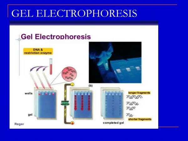
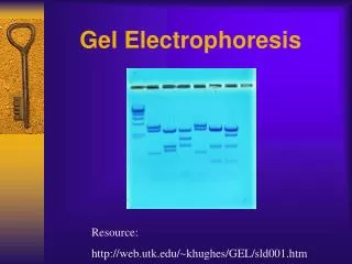
Separating the Fragments These DNA samples have already had restriction enzymes added- the DNA is in fragments now
Lighter fragments travel FURTHER from the starting well • A ladder is loaded into the first lane as a marker for fragment sizes • Identical DNA samples will have the same band # and locations