Case 2: Captive dik-dik
80 likes | 263 Vues
Case 2: Captive dik-dik. Kristen Baumann Christie Kranz. Madoqua guentheri smithi. African dik-dik Family Bovidae Small antelope found in Ethiopia, Kenya, Uganda, and Sudan 12-16 in at shoulder Elongated snout used for cooling Prefer areas that are over-grazed or disturbed.

Case 2: Captive dik-dik
E N D
Presentation Transcript
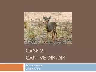
Case 2: Captive dik-dik Kristen Baumann Christie Kranz
Madoqua guentheri smithi • African dik-dik • Family Bovidae • Small antelope found in Ethiopia, Kenya, Uganda, and Sudan • 12-16 in at shoulder • Elongated snout used for cooling • Prefer areas that are over-grazed or disturbed
Protozoan Infections of African Ungulates • Toxoplasmosis • Babesiosis • African Trypanosomiasis (African Sleeping Sickness)
Case • 7 year old female dik-dik born in zoo • Lethargic, trembling, lacking motor coordination • Thin and dehydrated, labored breathing • Thoracic radiographs showed possible interstitial pneumonia • Blood collected, given antibiotics and vitamins • Became increasingly weaker, died next day
Necropsy • Lungs filled with fluid, covered with spots, bilaterally uniform • Enlarged lymph nodes, fat atrophied, small intestine was bleeding
Fig. 1. Lesions in the reticulum (A-C) of dik-dik. Foci of inflammation, large arrows. Protozoa in submucosa (small arrows) and mucosa (arrowheads). • Fig. 2. Lesions in lung. (A) Edema and infiltration of mononuclear cells in alveoli. (B) Several groups and individual protozoa (arrows). (C-D) Tissue cysts.