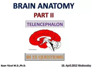
BRAIN ANATOMY PART II
490 likes | 770 Vues
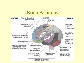
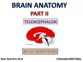
BRAIN ANATOMY PART II. TELENCEPHALON. IN 15 QUESTIONS. Kaan Yücel M.D.,Ph.D. 6.December .201 3 Friday. Learning Objectives Explain the components of the telencephalon Explain the boundaries , gyri , sulci in each lobe Explain the major Brodmann areas in each lobe

BRAIN ANATOMY PART II
E N D
Presentation Transcript
BRAIN ANATOMY PART II TELENCEPHALON IN 15 QUESTIONS Kaan Yücel M.D.,Ph.D. 6.December.2013Friday
Learning Objectives Explainthecomponents of thetelencephalon Explaintheboundaries, gyri, sulci in eachlobe ExplainthemajorBrodmannareasin eachlobe Explaintheventricularsystem Explainthefeatures of whitemattertracts
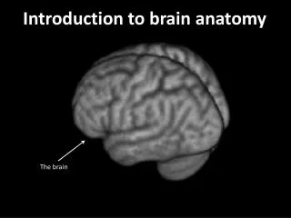
1. Telencephalon is composed of… Cerebralhemispheres Sulci, fissures, gyri, lobes partially separated by a deep longitudinal fissure, and which fill the area of the skull above the tentorium cerebelli and are subdivided into lobes based on their position. Basalganglia Insula Ventricularsystem Twolateralventricles, thirdventricle, fourthventricle White mattertracts Commissural, association, andprojectionfibres
2. Sulci & Gyri & Lobes Precentralgyrus
8. Brodmannareas Neocortex6 layers Paleocortex3-5 layers Archicortex3 layers 1909 Cytoarchitecture 47 functionalareas
8. Brodmannareas 4 8 3 5 7 6 9 1 10 46 39 46 19 10 2 4 4 4 5 43 41, 42 18 47 22 17 21 11 37 38
11. Fourthventricle FrontalLobe. ParietalLobe. OccipitalLobe. SeptumPellucidum. a. Rostrum of CorpusCallosum.b. Body of CorpusCallosum.c. Splenium of CorpusCallosum.10. Pons. 11.Medulla Oblongata. 12. Cerebellum. 13. SpinalCord. 14. Fourthventricle. 15. SinusConfluence.
12. Basalganglia Distributedset of brain structures in the telencephalon, diencephalon, and mesencephalon. The forebrain structures include : Caudate nucleus Putamen Nucleusaccumbens(or ventral striatum) Globuspallidus Corpus striatum
12. Basalganglia Caudatenucleus C-shaped structure closely associated with the lateral wall of the lateral ventricle. largest at its anterior pole (the head), and its size diminishes posteriorly as it follows the course of the lateral ventricle (the body) all the way to the temporal lobe (the tail), where it terminates at the amygdaloid nuclei.
12. Basalganglia Putamen- Caudateseparatedbyanterior limb of the internal capsule Connectedbybridges of cellsacrosstheinternalcapsule A striatedlook striatumorneostriatum Caudate+ Putamen=Striatum= Main recepient of afferentinput. Globuspallidus= Majorefferentoutputleavesfrom.
12. Basalganglia Third ventricle MRI of thebrain, T1-weighted axialcut.1, Putamen. 2, Pallidum. 3, Caudatenucleus. 4, Insula. 5, Lateralventricle. 6, Thalamus.
FunctionalAnatomy of theBasalGanglia Traditionally, the basal ganglia have been viewed as motor structures. It is only in the past 20 years that it has been recognized that these structures also may have a role in cognition and emotion.
FunctionalAnatomy of theBasalGanglia Alexander and his colleagues that the motor circuit is involved in ‘‘thecontrol of movementdirectionand in thescaling of movementamplitudeorvelocity’’ and ‘‘in theprogrammingandinitiation of internallygeneratedmovements.’’
FunctionalAnatomy of theBasalGanglia EXTRAPYRAMIDAL SYSTEM • GABA, dopamine, acetylcholine, and glutamine • Multiplepathways in thebasalgangliabothwith both excitatory and inhibitory functions. • Input to the basal ganglia is received from both thecerebral cortex and the thalamus. • Lesions in the basal ganglia result in uncoordinated and disorganized movement.
FunctionalAnatomy of theBasalGanglia motor - between additional motor area of the cerebral cortex and the lateral part of dorsal striatum – putamen automatic motor activity control of muscular tension initiating and fluent performing of motor actions executed by skeletal muscles especially during will dependent movements 2. oculomotor- between the frontal visual eye field of the cerebral cortex and the corpus of thecaudate (nucleus caudatus) belonging to the medial part of dorsal striatum
FunctionalAnatomy of theBasalGanglia 3. prefrontal (associative)- between dorso-lateral prefrontal cortex and the dorso-lateral part of the headof caudate (nucleus caudatus) (the frontal part of the medial part of dorsal striatum) choice of aims, planning, programming of the sequence of mental actions and behaviours, switching between sentences (the ability to change attitude flexibly), verbal and spatial working memory, selfcontrol and metacognition (self-consciousness)
FunctionalAnatomy of theBasalGanglia 4. latero-orbito-frontal- between lateral orbito-frontal cerebral cortex and the ventromedial part of the head of caudate (medial part of the dorsal striatum initiating social behaviours motivated by an award and in inhibiting behaviours, which can trigger punishment Dysfxn=> disinhibition
FunctionalAnatomy of theBasalGanglia 5. limbic (circuit of the anterior part of the cingulargyrus) - between the anterior part of the anterior cingulate gyrus and the ventral striatum (of which the main part is the nucleus accumbens). behavior control and adaptation of behaviours after making a mistake. responsible for correcting behavior following a mistake
12. Basalganglia Pangelinan MM, Zhang G, VanMeter JW, Clark JE, Hatfield BD, Haufler AJ. Beyond ageandgender: relationshipsbetweencorticalandsubcorticalbrainvolumeandcognitive-motor abilities in school-agechildren. Neuroimage. 2011 14;54(4):3093-3100. AlmeidaMontes LG, Ricardo-Garcell J, Barajas De La Torre LB, PradoAlcántara H, MartínezGarcía RB, Fernández-Bouzas A, AvilaAcosta D.Clinicalcorrelations of greymatterreductions in thecaudatenucleus of adultswithattentiondeficithyperactivitydisorder. J PsychiatryNeurosci. 2010;35(4):238-246. MitelmanSA, Canfield EL, Chu KW, Brickman AM, Shihabuddin L, Hazlett EA, Buchsbaum MS.Pooroutcome in chronicschizophrenia is associatedwithprogressiveloss of volume of theputamen.SchizophrRes. 2009;113(2-3):241-245.
12. Basalganglia MRI of thebrain, T1-weighted coronalcut.1, Lateral ventricle. 2, Caudate nucleus. 3, Putamen. 4, Temporal lobe (left side). 5, Sylvian fissure (lateral sulcus).
13. Commissuralfibers CORPUS CALLOSUM Freitag CM, Luders E, Hulst HE, Narr KL, Thompson PM, Toga AW, Krick C, Konrad C. Total brain volume and corpus callosum size in medication-naïve adolescents and young adults with autism spectrum disorder.Biol Psychiatry. 2009;66(4):316-319. Kitayama N, Brummer M, Hertz L, Quinn S, Kim Y, Bremner JD.Morphologic alterations in the corpus callosum in abuse-related posttraumatic stress disorder: a preliminary study.J NervMent Dis. 2007;195(12):1027-1209. Ballmaier M, Kumar A, Elderkin-Thompson V, Narr KL, Luders E, Thompson PM, Hojatkashani C, Pham D, Heinz A, Toga AW. Mapping callosal morphology in early- and late-onset elderly depression: an index of distinct changes in cortical connectivity. Neuropsychopharmacology. 2008;33(7):1528-1536. Black SE, Moffat SD, Yu DC, Parker J, Stanchev P, Bronskill M. Callosal atrophy correlates with temporal lobe volume and mental status in Alzheimer's disease.Can J Neurol Sci. 2000;27(3):204-209. VenkatasubramanianG, Jayakumar PN, Reddy VV, Reddy US, Gangadhar BN, Keshavan MS Corpuscallosumdeficits in antipsychotic-naïveschizophrenia: evidenceforneurodevelopmentalpathogenesis.PsychiatryRes. 2010;182(2):141-145.
Genu of corpuscallosum Forcepsminor Anteriorlimb of internalcapsule Septumpellucidum Caudatenucleus Putamen Globuspallidus Posteriorlimb of internalcapsule Thalamus Splenium of corpuscallosum Forcepsmajor
13. Commissuralfibers ANTERIOR COMMISSURE POSTERIOR COMMISSURE
15. Projectionfibers 1st neuron 2nd neuron 3rd neuron Internalcapsule Anteriorlimb [3] Posteriorlimb [8] Genu
Sensoryhomunculus Motor homunculus
Sensoryhomunculus Motor homunculus