Download
1 / 78
870 likes | 1.42k Vues
Anatomy of Brain. Dr. Rima Pathak. Gross Anatomy. Main Sulci & Gyri. Central. Cingulate. Parieto Occipital. Precentral gyrus. Premotor area 6. Postcentral gyrus. Cingulate. Superior frontal gyrus. Thalamus. Caudate. Infundibular stalk. Pineal gland. Mid brain. 4 th ventricle.

E N D
Anatomy of Brain Dr. Rima Pathak
Central Cingulate Parieto Occipital
Precentral gyrus Premotor area 6 Postcentral gyrus Cingulate Superior frontal gyrus Thalamus Caudate Infundibular stalk Pineal gland Mid brain 4th ventricle Sella Pons Inf Cerebellum medulla Spinal cord
Median longitudinal fissure Sup. Frontal Cingulate Inf. Frontal Sylvian Sup & middle Temporal
Cingulate gyrus Middle Frontal Gyrus Corpus callosum Inf Frontal Gyrus Septum pellucidum 3rd Ventricle Insular cortex Sup temporal gyrus Inf Temporal Gyrus hippocampus Internal Carotid A
lens Eye ball Optic nerve Ethmoid air cells Temporal horn of lat ventricle Nasopharynx Internal carotid artery Inf. Temporal gyrus Inf pons Vermis Formation of Basilar artery Parieto occipital sulcus Transverse Sinus
# Frontal Lobe Falx cerebri Caudate Thalamus & 3rd Ventricle Putamen Choroid plexus 4th Ventricle Splenium of corpus callosum
The Cerebral Hemispheres • Central sulcus(of Rolando) • Lateral sulcus(of Sylvius); • Parietooccipital sulcus; • Calcarine sulcus. • Preoccipital notch divide each cerebral hemisphere
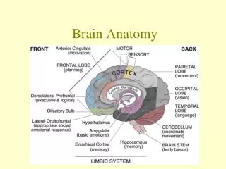
Frontal Lobe • Anterior tip of the brain (the frontal pole) to the central sulcus. • Inferiorly lateral sulcus/ Sylvian Fissure. • Medially extends anterior to an imaginary line from the top of the central sulcus to the corpus callosum.
Attention • Behavior, • Abstract thinking, • Problem solving, • Creative thought, • Emotion, • Intellect, • Initiative, • Judgment, • Coordinated movements, • Muscle movements, smell, • Physical reactions, • and Personality.
Broca's Area opercular and triangular sections of the inferior frontal gyrus. Expressive language, speech. Damage .Sparse Relevant Speech/Low word output.
Parietal Lobe • Anteriorly Central sulcus to an imaginary line connecting the top of the parietooccipital sulcus and the preoccipital notch. • Inferiorly it is bounded by the lateral sulcus and the imaginary continuation of this sulcus to the posterior boundary of the parietal lobe. • Medially surface, it is bounded posteriorly by the corpus callosum and calcarine sulcus, anteriorly by the frontal lobe • Posterior by the parietooccipital sulcus.
four anatomical boundaries: • the central sulcus • the parieto-occipital sulcus • the lateral sulcus (sylvian fissure) • the medial longitudinal fissure the two hemispheres.
Sensory cortex is located in the front part of the parietal lobe. Receives information from the spinal cord about the sense of touch, pressure, pain, and the perception of the position of body parts and their movements.
Fissure of Rolando (central sulcus) to the parieto-occipital fissure behind. Below, it is limited anteriorly by the fissure of Sylvius, while its posterior portion merges into the temporosphenoidal lobe.
Temporal lobe • Superiorly: to the lateral sulcus and the line forming the inferior boundary of the parietal lobe • Posteriorly: it extends to the line connecting the top of the parietooccipital sulcus and the preoccipital notch. • Medial surface: its posterior boundary is an imaginary line from the preoccipital notch to the splenium of the corpus callosum.
Functions include comprehension, naming, verbal memory and other language functions. Sound processing is controlled by the temporal lobes- in the Broca’s area and Wernicke’s area. • The underside (ventral) part high-level visual processing of complex stimuli such as faces (fusiform gyrus) and scenes (parahippocampal gyrus) object perception and recognition. • The medial temporal lobes (near the Sagittal plane that divides left and right cerebral hemispheres) are thought to be involved in episodic/declarative memory. • The hippocampi seem to be particularly important for memory function - particularly transference from short to long term memory and control of spatial memory and behavior.
Auditory & Visual memories, language, hearing & speech, language, behavior • Wernicke's Areasurrounds auditory cortex: • understanding & formulating speech. Damage Verbegeration/ Word salad.
Occipital lobe • Controls vision • homonomous vision loss from similarly positioned "field cuts" in each eye. • visual hallucinations. • parietal-temporal-occipital lesions color agnosia, movement agnosia, and agraphia.
Brainstem • Cerebrum Spinal cord • Motor and Sensory pathways • Cardiac and Respiratory • Reflexes • Originating in the brainstem • 10 of the 12 cranial nerves
Midbrian • center for ocular motion • The tectum (L. roof) of the midbrain consists of paired lumps, the superior and inferior colliculi (L. small hills). • This is dorsal to the cerebral aqueduct
Pons • coordinating eye and facial movements, facial sensation, hearing and balance. • This consists of: • A protruding basal portion, which is oval in the sagittal section; • And the overlying pontine tegmentum, which forms part of the floor of the 4th ventricle.
Medulla • breathing, blood pressure, heart rhythms and swallowing • reticular activating system is found in the midbrain, pons, medulla and part of the thalamus. It controls levels of wakefulness, enables people to pay attention to their environments, and is involved in sleep patterns • The point of attachment of most cranial nerves
Optic Pathway • Optic Chiasm is located beneath the hypothalamus and is where the optic nerve crosses over to the opposite side of the brain
Cingulate Gyrus • This is immediately superior to the corpus callosum. • it continues as the parahippocampal gyrus of the temporal lobe. • These 2 gyri give the appearance of encircling the diencephalon. • They, together with the olfactory bulb and tract, and certain other small cortical areas, are often referred to separately as the limbic lobe (L. limbus, border).
Pituitary Hypothalamus Complex • Hypothalamus is a region of the brain in partnership with the pituitary gland that controls the hormonal processes of the body as well as temperature, mood, hunger, and thirst. • This is inferior to the thalamus. • It is separated from it by the hypothalamic sulcus in the wall of the 3rd ventricle. • It also forms the floor of the ventricle. • Its inferior surface includes the infundibular stalk and the mammillary bodies. • The hypothalamus is the major visceral control centre of the brain. • It is involved in the limbic system as well.
Caudate • The round body bulging into the lateral ventricle and forming its floor is the caudate nucleus
Thalamus • located near the center of the brain and controls input and output to and from the brain • sensation of pain and attention.
borders on the 3rd ventricle. • major importance in both sensory and motor systems. • The line of attachment of the roof of this ventricle is marked by a horizontally oriented ridge, the stria medullaris thalami. • medial surface of the 2 thalami fuse in many brains called the interthalamic adhesion or massa intermedia. • Posteriorly, the thalamus protrudes over the most rostral part of the brainstem. • Anteriorly, it abuts the interventricular foramen. • No sensory information (except olfactory information) reaches the cerebral cortex without prior processing in the thalami. • anatomical loops characteristic of motor systems, typically involve the thalami .
Insular cortex • buried in the depths of the lateral sulcus. • It is concealed from view by portions of the frontal, parietal, and temporal lobes. • The portion of a given lobe overlying the insula is called an operculum (L. lid). • There are frontal, parietal, and temporal opercula.
Pineal Region • on top of the midbrain behind the thalamus • Pineal Gland controls the response to light and dark. • Its principal hormone melatonin, a derivative of amino acid tryptophan. stimulated by darkness and inhibited by light. • Follows circadian cycle with peak levels morning.
Short term memory to long term memory. • The Papez Circuit. • Amygdala controls emotions, social inhibitions, aggression
Cerebellum • lower back of the head and is connected to the brain stem. • It is the second largest structure of the brain and is made up of two hemispheres. • The cerebellum controls complex motor functions such as walking, balance, posture, and general motor coordination
Ventricular System • Four Ventricles of the brain are connected cavities within the brain, where cerebrospinal fluid is produced • cavity shown in the septum pellucidum is the so-called fifth ve.ntricle
Lateral Ventricles • Two ventricles enclosed in the cerebral hemispheres are called the lateral ventricles (first and second). They each communicate with the third ventricle through a separate opening called the Foramen of Munro
Third Ventricle • The third ventricle is in the center of the brain, and its walls are made up of the thalamus and hypothalamus. • The third ventricle connects with the fourth ventricle through a long tube called the Aqueduct of Sylvius.