Download
1 / 33
330 likes | 527 Vues
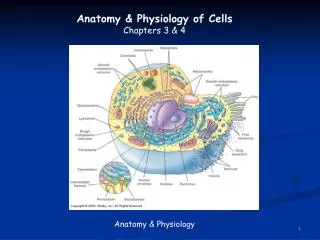
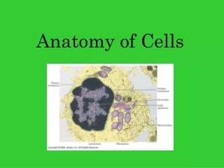
Chapter 3 Anatomy of Cells. Functional Anatomy of Cells. The typical cell (Figure 3-1) Also called composite cell Varies in size; all are microscopic (Table 3-1) Varies in structure and function (Table 3-2). Functional Anatomy of Cells. Cell structures

E N D
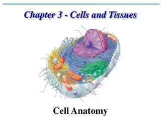
Functional Anatomy of Cells • The typical cell (Figure 3-1) • Also called composite cell • Varies in size; all are microscopic (Table 3-1) • Varies in structure and function (Table 3-2)
Functional Anatomy of Cells • Cell structures • Plasma membrane—separates the cell from its surrounding environment • Cytoplasm—thick gel-like substance inside of the cell composed of numerous organelles suspended in watery cytosol; each type of organelle is suited to perform particular functions (Figure 3-2) • Nucleus—large membranous structure near the center of the cell
Cell Membranes • Each cell contains a variety of membranes: • Plasma membrane (Figure 3-3) • Membranous organelles—sacs and canals made of the same material as the plasma membrane
Cell Membranes • Fluid mosaic model—theory explaining how cell membranes are constructed • Molecules of the cell membrane are arranged in a sheet • The mosaic of molecules is fluid; that is, the molecules are able to float around slowly • This model illustrates that the molecules of the cell membrane form a continuous sheet
Cell Membranes • Chemical attractions are the forces that hold membranes together • Groupings of membrane molecules form rafts, each of which float as a unit in the membrane (Figure 3-4) • Rafts may pinch inward, bringing material into the cell or organelle
Cell Membranes • Primary structure of a cell membrane is a double layer of phospholipid molecules • Heads are hydrophilic (water-loving) • Tails are hydrophobic (water-fearing) • Molecules arrange themselves in bilayers in water • Cholesterol molecules are scattered among the phospholipids to allow the membrane to function properly at body temperature • Most of the bilayer is hydrophobic; therefore water or water-soluble molecules do not pass through easily
Cell Membranes • Membrane proteins (Table 3-4) • A cell controls what moves through the membrane by means of membrane proteins embedded in the phospholipid bilayer • Some membrane proteins have carbohydrates attached to them, forming glycoproteins that act as identification markers • Some membrane proteins are receptors that react to specific chemicals, sometimes permitting a process called signal transduction
Cytoplasm and Organelles • Cytoplasm—gel-like internal substance of cells that includes many organelles suspended in watery intracellular fluid called cytosol
Cytoplasm and Organelles • Two major groups of organelles (Table 3-3): • Membranous organelles are specialized sacs or canals made of cell membranes • Nonmembranous organelles are made of microscopic filaments or other nonmembranous materials
Cytoplasm and Organelles • Endoplasmic reticulum (Figure 3-5) • Made of canals with membranous walls and flat, curving sacs arranged in parallel rows throughout the cytoplasm; extend from the plasma membrane to the nucleus • Proteins move through the canals
Cytoplasm and Organelles • Endoplasmic reticulum (cont.) • Two types of endoplasmic reticulum: • Rough endoplasmic reticulum • Ribosomes dot the outer surface of the membranous walls • Ribosomes synthesize proteins, which move toward the Golgi apparatus and then eventually leave the cell • Function in protein synthesis and intracellular transportation
Cytoplasm and Organelles • Two types of endoplasmic reticulum (cont.) • Smooth endoplasmic reticulum • No ribosomes border membranous wall • Functions are less well established and probably more varied than for rough endoplasmic reticulum • Synthesizes certain lipids and carbohydrates and creates membranes for use throughout cell • Removes and stores Ca++ from cell’s interior.
Cytoplasm and Organelles • Ribosomes (Figure 3-6) • Many are attached to the rough endoplasmic reticulum and many lie free, scattered through the cytoplasm • Each ribosome is a nonmembranous structure made of two pieces, a large subunit and a small subunit; each subunit is composed of rRNA • Ribosomes in the endoplasmic reticulum make proteins for “export” or to be embedded in the plasma membrane; free ribosomes make proteins for the cell’s domestic use
Cytoplasm and Organelles • Golgi apparatus • Membranous organelle consisting of cisternae stacked on one another and located near the nucleus (Figure 3-7) • Processes protein molecules from the endoplasmic reticulum (Figure 3-8) • Processed proteins leave the final cisterna in a vesicle; contents may then be secreted to outside the cell
Cytoplasm and Organelles • Lysosomes (Figure 3-9) • Made of microscopic membranous sacs that have “pinched off” from Golgi apparatus • The cell’s own digestive system; enzymes in lysosomes digest the protein structures of defective cell parts, including plasma membrane proteins, and particles that have become trapped in the cell
Cytoplasm and Organelles • Proteasomes (Figure 3-10) • Hollow, protein cylinders found throughout the cytoplasm • Break down abnormal/misfolded proteins and normal proteins no longer needed by the cell • Break down protein molecules one at a time by tagging each one with a chain of ubiquitin molecules and unfolding it as it enters the proteasome, then breaking apart peptide bonds
Cytoplasm and Organelles • Peroxisomes • Small membranous sacs containing enzymes that detoxify harmful substances that enter the cells • Often seen in kidney and liver cells
Cytoplasm and Organelles • Mitochondria (Figure 3-11) • Made up of microscopic sacs; wall composed of inner and outer membranes separated by fluid; thousands of particles make up enzyme molecules attached to both membranes • The “power plants” of cells; mitochondrial enzymes catalyze series of oxidation reactions that provide about 95% of cell’s energy supply • Each mitochondrion has a DNA molecule, allowing it to produce its own enzymes and replicate copies of itself
Nucleus • Definition—spherical body in center of cell; enclosed by an envelope with many pores
Nucleus • Structure • Consists of nuclear envelope (composed of two membranes each with essentially the same molecular structure as plasma membrane) surrounding nucleoplasm; nuclear envelope has holes called nuclear pores (Figure 3-12)
Nucleus • Structure (cont.) • Contains DNA (heredity molecules), which appear as the following: • Chromatin threads or granules in nondividing cells • Chromosomes in early stages of cell division • Functions of nucleus are functions of DNA molecules; DNA determines both structure and function of cells and heredity
Cytoskeleton • The cell’s internal supporting framework made up of rigid, rodlike pieces that provide support and allow movement and mechanisms that can move the cell or its parts (Figure 3-13)
Cytoskeleton • Cell fibers • Intricately arranged fibers of varying lengths that form a three-dimensional, irregularly shaped lattice • Fibers appear to support the endoplasmic reticulum, mitochondria, and “free” ribosomes
Cytoskeleton • Cell fibers (cont.) • Smallest cell fibers are microfilaments (Figure 3-14) • “Cellular muscles” • Made of thin, twisted strands of protein molecules that lie parallel to the long axis of the cell • Microfilaments can slide past each other, causing shortening of the cell
Cytoskeleton • Cell fibers (cont.) • Intermediate filaments are twisted protein strands slightly thicker than microfilaments; they form much of the supporting framework in many types of cells • Microtubules are tiny, hollow tubes that are the thickest of the cell fibers; they are made of protein subunits arranged in a spiral fashion; their function is to move things around in the cell
Cytoskeleton • Centrosome • An area of the cytoplasm near the nucleus that coordinates the building and breaking of microtubules in the cell • Nonmembranous structure also called the microtubule-organizing center (MTOC) • Plays an important role during cell division • The general location of the centrosome is identified by the centrioles
Cytoskeleton • Cell extensions • Cytoskeleton forms projections that extend the plasma membrane outward to form tiny, fingerlike processes
Cytoskeleton • There are three types of these processes; each has specific functions (Figure 3-15): • Microvilli—found in epithelial cells that line the intestines and other areas where absorption is important; they help to increase the surface area manyfold • Cilia and flagella—cell processes that have cylinders made of microtubules at their core; cilia are shorter and more numerous than flagella; flagella are found only on human sperm cells
Cell Connections • Cells are held together by fibrous nets that surround groups of cells (e.g., muscle cells), or cells have direct connections to each other • There are three types of direct cell connections (Figure 3-16)
Cell Connections • Desmosome • Fibers on the outer surface of each desmosome interlock with each other; anchored internally by intermediate filaments of the cytoskeleton • Spot desmosomes, connecting adjacent membranes, are like “spot welds” at various points • Belt desmosomes encircle the entire cell like a collar
Cell Connections • Gap junctions—membrane channels of adjacent plasma membranes adhere to each other; have two effects: • Form gaps or “tunnels” that join the cytoplasm of two cells • Fuse two plasma membranes into a single structure
Cell Connections • Tight junctions • Occur in cells that are joined by “collars” of tightly fused material • Molecules cannot permeate the cracks of tight junctions • Occur in the lining of the intestines and other parts of the body, where it is important to control what gets through a sheet of cells