Download
1 / 28
280 likes | 306 Vues
Explore the structure and function of typical cells compared to reality, including components like plasma membrane, cytoplasm, organelles, and nucleus. Learn about specialized cells like nerve cells, muscle cells, and RBCs, with detailed insights on cell extensions and the cytoskeleton. Discover the intricate world of cells in this informative guide.

E N D
“Typical” Cell vs. Reality • “Typical” cells do not actually exist in the body • Cell are specialized structure to carry out specialized functions (see Table 3-1, pg. 73) • Nerve cells: have long extension transmit nerve impulses throughout the body • Muscle cells: contain fibers that slide together contracts/movement of body parts • RBCs: contain hemoglobin carry oxygen from lungs to various parts of the body
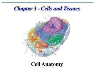
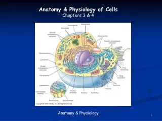
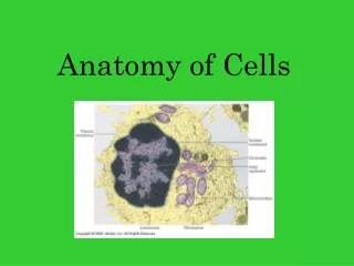
Main Components of Cell Structure • Plasma Membrane • Cytoplasm • Cytosol (intracellular fluid) • Organelles • Nucleus • Cytoskeleton • Cell’s internal supporting framework
Plasma Membrane • Phospholipid bilayer • Hydrophilic heads • Hydrophobic tails • Majority of membrane is hydrophobic – water and water-soluble molecules cannot pass • Cholesterol – steroid lipid; provides stabilization for the plasma membrane
Plasma Membrane • Embedded proteins • Penetrate into the hydrophobic regions of the plasma membrane • Transport mechanism • Transport proteins are often specific for certain molecules • “Gates” can open or close
Plasma Membrane • Peripheral Proteins • Glycoproteins (carbohydrates and proteins) • Identification markers • Recognize self vs. non-self (immune system) • Receptors • Proteins that react in the presence of hormones or other regulatory chemicals • Trigger metabolic changes within the cells • Signal transduction
Cytoplasm • Cytosol • Watery solution • Intercellular fluid (ICF) • Organelles • “tiny organs” • Thicken the cytosol to a gel-like consistency
Endoplasmic Reticulum • Rough ER • Presence of ribosomes • Protein synthesis • Intracellular transport • Smooth ER • Lipid and carbohydrate synthesis • Replenishes cell membrane material http://micro.magnet.fsu.edu/cells/endoplasmicreticulum/images/endoplasmicreticulumfigure1.jpg
Ribosomes • Attached to Rough ER or scattered throughout cytoplasm • Composed of a large and a small subunit • Each subunit contains RNA (rRNA) bonded to protein • Function – Protein synthesis • Cell’s “protein factory”
Golgi Apparatus • Consists of tiny sacs or cisternea • “processing & packaging plant” • Export proteins make in the Rough ER out of the cell • Secretion • Fig 3-5, page 81
Lysosomes • Vesicles that have pinched off from the Golgi apparatus • Contain enzymes capable of breaking down cell components • “digestive bags” or “cellular garbage disposals” • Ex: rid cells of bacteria; Scavenger WBCs
Peroxisomes • Similar to lysosomes • Small sac containing enzymes • Important in kidney and liver cells • Detoxification functions in the body
Mitochondria • Structure: • Two membranes (sac within a sac) • Inner membrane contains folds (cristae) • Function: • Enzymes embedded in cristae – essential in making adenosine tri-phosphate (ATP) • Cell “power plant” • # of mitochondria based on amt of work done by cell • Ex: liver cells > sperm cells • Self-replicating – based on energy needs • Aerobic exercise increases # of mitochondria in skeletal muscle cells
Nucleus • Large, spherical organelle • Enclosed by a two nuclear membranes = nuclear envelope • Nuclear pores – selectively allow molecules to enter/leave nucleus • Contains DNA (genetic information) • Chromatin – uncondensed genetic material • Chromosomes – condensed genetic material • Nucleolus – synthesizes rRNA
Cytoskeleton • Cell Fibers • Microfilaments • “Cellular muscles” • Thin, twisted strains of protein • Can contract (ex: muscle cells) • Intermediate Filaments • slightly thicker • Main component of the supporting framework in many cell types • Microtubules • Thickest of the cell fibers; tiny, hollow tubes • Cell “engine” – help with movement within the cell and the cell itself
Cytoskeleton • Centrosome • Located near the nucleus • “microtubule-organization center” • Important role in cell division – move chromosomes around the cell • Centrioles • Cylindrical structures within the centrosome • Replicate prior to cell division • Roles in cell division
Cell Extensions • Microvilli • Epithelial cells found where absorption is necessary (ex: small intestine) • Increase surface area • Cilia • Transport fluid across a cell surface • Ex: Line the respiratory tract – move mucous upward • Ex: Assist the ovum to move towards the uterus • Flagella • Single, long structures; aids in locomotion • Ex: sperm cells
Anthony’s Textbook of Anatomy and Physiology 17th Edition. Thibodeau, Gary A. PhD and Patton, Kevin T. PhD. Mosby, Inc.