Download
1 / 44
550 likes | 1.47k Vues
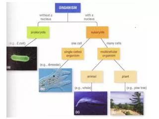
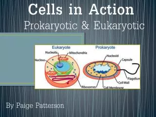
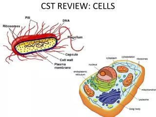
Chapter 4: Functional Anatomy of Prokaryotic and Eukaryotic Cells. Prokaryote Eukaryote. True nucleus. Pre-nucleus. One circular chromosome, not in a membrane No histones No membrane-bound organelles Complex cell walls (peptidoglycan in bacteria) Binary fission.

E N D
Chapter 4: Functional Anatomy of Prokaryotic and Eukaryotic Cells
Prokaryote Eukaryote True nucleus Pre-nucleus • One circular chromosome, not in a membrane • No histones • No membrane-bound organelles • Complex cell walls (peptidoglycan in bacteria) • Binary fission • Paired chromosomes, in nuclear membrane • Histones bound to DNA • Membrane-bound organelles • Simpler cell walls (polysaccharide) • Mitosis
Prokaryotic Cells: Morphology • Average size: 0.2 -2.0 µm in diameter 2.0-8.0 µm in length • Basic shapes/morphologies: Coccus (spherical) Bacillus* (rod-shaped) Spiral Coccobacillus
Prokaryotic cells: Arrangements Pairs: diplo-diplococci diplobacilli Chains: strepto- streptococci streptobacilli Clusters: staphylo-staphylococci
Prokaryotic cells: Structures external to the cell wall
Prokaryotic cell structures:Glycocalyx • Capsule: Glycocalyx firmly attached to the cell wall • Sticky outer coat • Polysaccharide and/or polypeptide composition • Aids in attachment of cell to a substrate • Helps prevent dehydration • Impairs phagocytosis (host defense mechanism) Figure 4.6a http://lecturer.ukdw.ac.id/dhira/BacterialStructure/SurfaceStructs.html
Prokaryotic cell structures:Flagella • Cellular propellers • Three basic parts • Filament: Made of chains of flagellin protein • Hook • Basal body: anchor to the cell wall and membrane • Rotation in the basal body propels the cell Figure 4.8
Flagella Arrangement Prokaryotic cell structures: Flagella ( flagella at both poles) (flagella distributed over the entire cell) (tuft from one pole) Figure 4.7
Prokaryotic cell structures: FlagellaMotility Taxis: movement toward or away from a stimulus -chemotaxis -phototaxis Figure 4.9
Prokaryotic cell structures:Flagella Motile E. coli with flourescently-labelled flagella from the Roland Institute at Harvard
Prokaryotic cell structures:Axial Filaments • Axial filaments= endoflagella • Spirochete movement • Anchored at one end of a cell • Contained within an outer sheath • Rotation causes cell to move • Spiral/corkscrew motion Figure 4.10a
Prokaryotic cell structures:Fimbriae and Pili • Composed of protein pilin • Fimbriae allow attachment to surfaces/other cells • Few to hundreds per cell • Pili are used to transfer DNA from one cell to another • Longer, 1-2 per cell Figure 4.11
Prokaryotic cells: The cell wall
Prokaryotic cell structures:Cell Wall • Main functions of the cell wall • Prevents osmotic lysis • Helps maintain cell shape • Point of anchorage for flagella • Contains peptidoglycan (in bacteria)
Prokaryotic cell structures: Cell WallPeptidoglycan (or murein) Macromolecular network: • Polymer of a repeating disaccharide unit (backbone) • Backbones linked by polypeptides Figure 4.13a
Prokaryotic cell structures:Gram-positive Cell Walls Gram-positive cell wall • ThickPG layer; rigid structure • Teichoic acids: • Lipoteichoic acid links PG to plasma membrane • Wall teichoic acid links PG sheets • Polysaccharides & teichoic acids are cellular antigens
Prokaryotic cell structures:Gram-negative Cell Walls Gram-negative cell wall • ThinPG layer • Surrounded by an outer membrane • Protection from phagocytes, some antibiotics • Periplasm (contains PG, degradative enzymes, toxins) • Lipopolysaccharide (LPS) • O polysaccharide antigen • Lipid A is called endotoxin • Porins (proteins) form channels through membrane Figure 4.13b, c
Prokaryotic cell structures:Cell Walls Gram-positive cell walls Gram-negative cell walls • Thin peptidoglycan • No teichoic acids • Outer membrane • Lipopolysaccharide • Porins • Thick peptidoglycan • Teichoic acids
Gram stain mechanism • Step 1: Crystal violet (primary stain) • Enters the cytoplasm (stains) both cell types • Step 2: Iodine (mordant) • Forms crystals with crystal violet (CV-I complexes) that are too large to diffuse across the cell wall • Step 3: Alcohol wash (decolorizer) • Gm +: dehydrates PG, making it more impermeable • Gm -: dissolves outer membrane and pokes small holes in thin PG through which CV-I can escape • Step 4: Safranin (counterstain) • Contrasting color to crystal violet
Prokaryotic cell structures: Cell WallAtypical Cell Walls • Mycoplasma • Smallest known bacteria • Lack cell walls • Sterols in plasma membrane protect from lysis • Acid-fast cells (Mycobacterium and Nocardia genera) • Mycolic acid (waxy lipid) layer outside of PG resists typical dye uptake • Archaea • Wall-less, or • Walls of pseudomurein (altered polysaccharide and polypeptide composition vs. PG)
Prokaryotic cell structures: Cell WallDamage to Cell Walls • Lysozyme attacks disaccharide bonds in peptidoglycan • Penicillin inhibits peptide cross-bridge formation in peptidoglycan • Bacteria with weakened cell walls are very susceptible to osmotic lysis
Prokaryotic cells: Structures internal to the cell wall
Prokaryotic cell structures:Plasma Membrane • Phospholipid bilayer • Proteins • Fluid Mosaic Model • Membrane is as viscous as olive oil • Proteins move to function • Phospholipids rotate and move laterally Figure 4.14b
Prokaryotic cell structures:Plasma Membrane Main function: selective barrier • Selective permeability allows passage of select molecules • Small molecules (H2O, O2, CO2, some sugars) • Lipid-soluble molecules (nonpolar organic)
Prokaryotic cell structures:Plasma Membrane Movement across membranes: Passive Processes • Passive processes do not require energy (ATP)—involves movement down a concentration gradient • Simple diffusion: Movement of a solute from an area of high concentration to an area of low concentration • Facilitated diffusion: Solute combines with a transporter protein to move down its gradient • Osmosis: Diffusion of water down its gradient • Diffusion continues until the solute is evenly distributed (state of equilibrium)
Osmosis: Movement of water across a selectively permeable membrane from an area of higher water concentration to an area of lower water concentration Water moving down its concentration gradient Prokaryotic cell structures:Plasma Membrane Figure 4.18
Prokaryotic cell structures:Plasma Membrane Three types of osmotic solutions: Hypotonic solution* Isotonic solution Hypertonic solution [sol]solution = [sol]cell [sol]solution < [sol]cell [sol]solution > [sol]cell Osmotic lysis Plasmolysis [sol]cell = concentration of solute inside the cell Figure 4.18c-e
Prokaryotic cell structures:Plasma Membrane Movement across membranes: Active Transport Processes • Active transport of substances requires a transporter protein and ATP • Solute movement up/against its concentrationgradient • Important when nutrients are scarce
Prokaryotic cell structures:Other Internal Structures • Cytoplasm: substance inside plasma membrane • 80% water, contains mainly proteins, carbohydrates, lipids, inorganic ions and low-molecular weight compounds • Chromosomal DNA: single circular string of double-stranded DNA attached to the plasma membrane • Located in the “nucleoid” area of the cell • Plasmids: small, circular, extrachromosomal DNA • Genes encoded typically not required for survival under normal conditions
Prokaryotic cell structures:Other Internal Structures • Ribosomes: machinery for protein synthesis (translation) • Tens of thousands of ribosomes per cell Complete 70S ribosome
Prokaryotic cell structures:Other Internal Structures Inclusions: Reserve deposits • Contents: • Phosphate reserves (for ATP generation) • Energy reserves • Energy reserves • Energy reserves • Ribulose 1,5-diphosphate carboxylase for CO2 fixation • Protein covered cylinders • Iron oxide (destroys H2O2) Type: • Metachromatic granules (volutin) • Polysaccharide granules • Lipid inclusions • Sulfur granules • Carboxysomes • Gas vacuoles • Magnetosomes
Prokaryotic cell structures:Other Internal Structures Endospores • Specialized resting cells (metabolically inactive) • Resistant to desiccation, heat, chemicals • Can remain dormant for thousands of years • Bacillus, Clostridium genera only (Gram-positive) • Sporulation: Endospore formation • Triggered by deficiency of a key nutrient • Germination: Return to vegetative state • Triggered by physical/chemical damage to endospore’s coat
Stained pus from a mixed anaerobic infection http://qlab.com.au www.textbookofbacteriology.net
Functional Anatomy of Eukaryotic Cells Figure 4.22
Functional Anatomy of Eukaryotic Cells:Flagella and Cilia • Contain cytoplasm, enclosed by plasma membrane • Move in a wavelike motion Figure 4.23
Functional Anatomy of Eukaryotic Cells:Flagella and Cilia Figure 4.23a, b
Functional Anatomy of Eukaryotic Cells:Cell Walls and Glycocalyx • Cell walls • Plants, algae, fungi • Polysaccharide; simpler than prokaryotic cell walls • Glycocalyx • Carbohydrates extending from animal plasma membrane • Anchored to cell membrane • Strengthens surface, aids in attachment
Similar to prokaryotic PM structure and function: Phospholipid bilayer Peripheral proteins Integral proteins EXCEPT, they contain Sterols (complex lipids, help strengthen membrane) Site for endocytosis (phagocytosis and pinocytosis) Selective permeability Passive transport processes Active transport processes Functional Anatomy of Eukaryotic Cells:Plasma Membrane
Functional Anatomy of Eukaryotic Cells:Cytoplasm • Cytoplasm Substance inside plasma membrane and outside nucleus • Cytoskeleton Microfilaments, intermediate filaments, microtubules • Contains organelles Specialized functions (nucleus, ER, Golgi complex, mitochondria, lysosomes, ribosomes)
Functional Anatomy of Eukaryotic Cells:Ribosomes • Larger, denser than prokaryotic • Eukaryotic ribosomes: 80S Prokaryotic ribosomes: 70S • Membrane-bound pool (attached to ER) • Free pool (in cytoplasm)
Functional Anatomy of Eukaryotic Cells:Nucleus • Storage for almost whole cell’s genetic information • Enclosed by nuclear envelope • Proteins bound to DNA (histones and nonhistones) Figure 4.24
Contains DNA and ribosomes (70S) Double membrane Inner membrane folds: cristae Portion inside of inner membrane: matrix Proteins involved in respiration and ATP production in cristae and matrix Chloroplasts: enzymes for light-gathering necessary for photosynthesis Functional Anatomy of Eukaryotic Cells: Mitochondrion Figure 4.27
Endosymbiotic Theory • Theory on evolution of eukaryotic cells’ organelles • Prokaryotes: 3.5-4 byo • Eukaryotes: ~2.5 byo • Mitochondria and chloroplasts share properties with prokaryotes • Reproduce independently of cell • Size/shape • Circular DNA • Their ribosomes are 70S